10-E Farmfield Avenue, Charleston, SC 29407

235-213-0595

Author: Dr. O’Neal

  • Do You Need Antibiotics Before Dental Treatment?

    Do You Need Antibiotics Before Dental Treatment?

    Far fewer people need antibiotics before dental treatment than commonly believe they do. The guidance narrowed considerably about twenty years ago, and a lot of patients are still following advice given before it changed.

    Here is who still needs premedication, who no longer does, and why taking it unnecessarily is not a harmless precaution.

    Key takeaways

    • Only a small, defined group at highest risk of infective endocarditis still needs cover.
    • Prosthetic joints generally do not require premedication any more.
    • Routine fillings, cleanings and most root canal treatment need nothing for most patients.
    • The decision belongs to your physician or surgeon, not to us and not to a search result.

    What premedication is actually for

    Dental procedures can release oral bacteria into the bloodstream briefly. In almost everyone that is harmless and resolves within minutes.

    In a small number of people, those bacteria can settle on abnormal heart tissue and cause infective endocarditis — rare, but serious. A single antibiotic dose an hour beforehand is intended to reduce that risk in exactly those patients, and no one else.

    Who still needs it

    Current position by condition
    ConditionPremedication?Note
    Prosthetic heart valve, or prosthetic repair materialYesHighest-risk category
    Previous infective endocarditisYesRecurrence risk
    Certain congenital heart conditionsYesUnrepaired cyanotic disease and specific repairs
    Cardiac transplant with valve problemsYesTransplant recipients who develop valvulopathy
    Hip or knee replacementGenerally noNot recommended routinely since 2015
    Heart murmur, stents, bypass, pacemakerNoNot in the current categories
    Mitral valve prolapseNoRemoved from guidance in 2007

    Swipe the table sideways to see every column.
    Only the highest-risk cardiac categories are still covered
    Only the highest-risk cardiac categories remain covered by current guidance.

    Two entries there account for most of the confusion. Mitral valve prolapse and straightforward heart murmurs were dropped in 2007, and prosthetic joints in 2015 — but a lot of patients were told once and never told again.

    What to do if you are uncertain

    The safest position when your history is unclear is neither to take antibiotics by default nor to skip them by default, but to establish the answer once and record it.

    Ask the physician who manages the underlying condition — the cardiologist, or the surgeon who placed the joint — to confirm in writing whether cover is required. That letter then travels with you to every dental appointment, and nobody has to guess again.

    Why the guidance narrowed

    It was not a cost-saving exercise. The evidence simply did not support the breadth of the old recommendations.
    Unnecessary prophylaxis carries real costs of its own
    Unnecessary prophylaxis carries real costs of its own.

    Bacteria enter the bloodstream during ordinary chewing and toothbrushing too, far more often across a year than during occasional dental visits. Against that background, single-dose cover before a dental appointment prevents very few cases — while allergic reactions, gastrointestinal effects and antibiotic resistance are real and cumulative. The current position is that good oral health and regular dental care protect these patients more than prophylaxis does.

    Joint replacements specifically

    This is the most common source of disagreement, and the honest position is that guidance and individual practice do not always match.
    Routine cover for prosthetic joints is no longer recommended
    Routine cover for prosthetic joints is no longer recommended.

    The ADA guideline is clear that antibiotics are not generally recommended before dental procedures for patients with prosthetic joints. Some orthopaedic surgeons still advise cover for particular patients — recent surgery, immunosuppression, previous joint infection — and where your surgeon has done so, we follow it. What we will not do is start it on our own initiative.

    What we need from you

    1. Tell us at booking, not on the day. Premedication is taken about an hour beforehand — discovering the need on arrival means rescheduling.
    2. Bring the instruction if you have one. A letter or note from your cardiologist or surgeon settles it immediately.
    3. Bring a current medication list, including anything over the counter.
    4. Tell us about allergies, particularly to penicillin — alternatives exist and are used routinely.
    5. If you are unsure, say so. We would rather check with your physician than guess in either direction.

    Bring your medication list and any letter from your physician
    Bring your medication list and any written instruction from your physician.

    Which procedures it applies to

    Even for patients who do need cover, it is not required for everything. The distinction is whether the procedure involves manipulating gum tissue or the region around the root tip.

    Procedures that do and do not require cover, for patients in a listed category
    ProcedureCover needed?
    Root canal treatment involving the root tipYes
    ExtractionYes
    Scaling and periodontal treatmentYes
    Apicoectomy and other endodontic surgeryYes
    Routine fillings above the gumlineNo
    Radiographs, impressions, fitting a crownNo
    Local anaesthetic in uninfected tissueNo

    Swipe the table sideways to see every column.

    If you are unsure whether your appointment falls on the left or right of that line, ask when you book and we will tell you.

    If you are allergic to penicillin

    Amoxicillin is the usual first choice, but a documented penicillin allergy is not a barrier — several established alternatives exist and are used routinely.

    It is worth knowing that most people labelled penicillin-allergic turn out not to be when formally tested, often because a childhood rash was attributed to the drug. That is a conversation for your physician rather than something to resolve in a dental chair, but it is worth having, because the alternatives are generally less effective.

    What about an active infection?

    This is a different question and often confused with the one above. Premedication prevents a theoretical problem in a healthy mouth; antibiotics for an active infection treat one that already exists.

    When guidance is unclear, the prescribing decision sits with your physician
    Where guidance is unclear, the prescribing decision sits with your physician.

    Where there is swelling or spreading infection we prescribe antibiotics as part of treatment, regardless of your cardiac or joint history. But as our abscess guide explains, antibiotics never resolve a dental infection on their own — the source has to be removed.

    Tell us at booking, not on the day — premedication is timed to the appointment
    Tell us when you book, so the timing works.

    If you have been premedicating for years and nobody has revisited it, it is worth asking at your next medical appointment. What to expect at your first visit covers the medical history we take, Medication history changes endodontic planning in several other ways too, as root canals later in life sets out. and the ADA’s summary of antibiotic prophylaxis before dental procedures sets out the current position and its sources.

    Frequently asked questions

    I had premedication years ago. Do I still need it?

    Quite possibly not. The guidance narrowed substantially in 2007 and again for joint replacements in 2015, so a great many people who once took antibiotics before dental visits no longer need to. Do not stop on the strength of a web page — ask the physician who advised it.

    Do I need antibiotics because of my knee or hip replacement?

    In general, no. The ADA guideline states prophylactic antibiotics are not recommended before dental procedures for patients with prosthetic joints. Some surgeons still advise it for individual patients, and that decision is theirs.

    Which heart conditions still require it?

    A small group at highest risk of an adverse outcome: prosthetic heart valves or prosthetic material used in valve repair, previous infective endocarditis, certain congenital heart conditions, and cardiac transplant recipients with valve problems.

    Who decides — you or my doctor?

    Your physician or surgeon. We follow their instruction and record it. If nobody has advised premedication and you are not in a listed category, none is needed.

    Have Questions or Ready to Schedule?

    Our team is here to help — call, email, or request an appointment online.
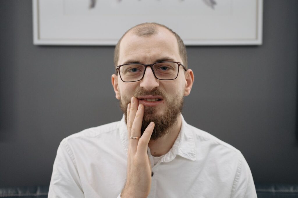
    About this guide. Written for patients of Southeast Endodontics, PC, Charleston, SC. It summarises published guidance and is not medical advice — never start or stop premedication on the strength of a web page. That decision belongs to your physician.

  • Root Canal While Pregnant: Is It Safe?

    Root Canal While Pregnant: Is It Safe?

    Being told you need a root canal while pregnant sets off an entirely reasonable set of worries about anaesthetic, X-rays and timing. The short answer is that treatment is safe — and that delaying an infection is the genuinely risky option.

    Here is what the professional guidance actually says, how we adapt the appointment, and the one situation where waiting is reasonable.

    Key takeaways

    • The ADA and ACOG both support necessary dental treatment during pregnancy.
    • The second trimester is the comfortable window for anything that can be planned.
    • Dental X-rays with shielding deliver an extremely small dose.
    • An untreated infection is a systemic risk to you, which makes it a risk to the pregnancy.

    Why treating is safer than waiting

    The instinct to postpone everything until after delivery is understandable, and for elective work it is often right. An active dental infection is not elective work.

    An untreated dental infection is a systemic risk, not a local one
    An untreated dental infection is a systemic problem, not a local one.

    An infected tooth is a source of bacteria and inflammation that does not stay local — it can spread into the tissues of the face and neck, and it brings pain, disrupted sleep and difficulty eating with it. None of that is neutral during pregnancy. Both ACOG and the ADA are explicit that treatment for teeth needing prompt attention should not be delayed.

    Timing, when there is a choice

    Dental treatment by trimester
    StagePlanned treatmentWhy
    First trimesterDefer if it can safely waitOrgan development; nausea often at its worst
    Second trimesterIdeal windowOrgan development complete, lying back still comfortable
    Third trimesterPossible, with adjustmentsLying flat becomes uncomfortable; shorter visits
    Any trimesterActive infection or severe painTreat now — delay is the greater risk

    Swipe the table sideways to see every column.

    The second trimester is the comfortable window for planned treatment
    The second trimester is the comfortable window for planned treatment.

    If you have facial swelling, fever, or pain that is keeping you awake, none of the above applies — that is same-day care regardless of stage. Our emergency guide covers what counts.

    X-rays: the question we get most

    Dental radiographs are among the lowest-dose imaging in medicine, and the beam is directed at the jaw rather than the abdomen. With a lead apron and thyroid collar, exposure to the uterus is negligible.

    Lead apron and thyroid collar; the dose is extremely small either way
    Lead apron and thyroid collar, and only the images needed to treat the tooth safely.

    The alternative is worse. Treating a root canal without seeing the root anatomy is how canals get missed, which is how treatment fails — see why root canals fail. We take the minimum needed, not a routine full series.

    Anaesthetic and medication

    Local anaesthetic is not only safe but important. Adequate numbing means less pain and less stress, both of which matter more during pregnancy, not less.

    Lidocaine at standard dental doses is well established in pregnancy
    Lidocaine at standard dental doses is well established in pregnancy.
    Medication during pregnancy: what changes
    ItemUsual positionNote
    Local anaesthetic (lidocaine)Used as normalWell established at standard dental doses
    Paracetamol / acetaminophenFirst-line for painPreferred analgesic in pregnancy
    Ibuprofen and other NSAIDsGenerally avoidedParticularly in the third trimester — ask your obstetrician
    Antibiotics, if neededSelected carefullySeveral are routinely used; some are avoided
    Nitrous oxide sedationUsually deferredWe would normally avoid it and manage anxiety differently

    Swipe the table sideways to see every column.

    That table is a guide, not a prescription. Tell us what you are already taking and who is looking after your pregnancy, and we work within their advice — our page on managing dental anxiety covers the non-drug approaches, which matter more here than usual.

    How the appointment itself changes

    Practical adjustments do most of the work. Later in pregnancy, lying flat can compress the vena cava and make you feel faint, so we tilt the chair slightly to the left and support you with a cushion.

    Shorter appointments and a slight left tilt make later visits easier
    Shorter appointments and a slight left tilt make later visits considerably easier.

    We also keep appointments shorter, build in breaks, and schedule for the time of day your nausea is least troublesome. None of this is complicated — it simply needs knowing in advance, so tell us when you book rather than on the day.

    Why dental problems surface during pregnancy

    It is not coincidence that so many people meet their first serious dental problem while pregnant.

    Hormonal change makes gums more reactive to plaque, producing the swelling and bleeding known as pregnancy gingivitis in a large proportion of pregnancies. Morning sickness adds stomach acid to the enamel, and cravings often mean more frequent snacking. None of that causes a root canal on its own, but together they accelerate problems that were already developing.

    One practical tip: after vomiting, rinse with water or a fluoride mouthwash rather than brushing immediately. Enamel is temporarily softened by the acid, and brushing straight away scrubs some of it away.

    Coordinating with your obstetrician

    For a straightforward root canal in an uncomplicated pregnancy, no clearance is needed. Where there are complications — a high-risk pregnancy, medication that affects bleeding, or an existing condition — we speak to your obstetrician first.

    We coordinate with your obstetrician where anything is uncertain
    Where anything is uncertain, we work to your obstetrician’s advice.

    That is a short conversation, not a delay. Bring their contact details to the appointment and we will handle it.

    What we will not do

    Being clear about the limits is as useful as listing what is safe.

    Elective work waits. Whitening, cosmetic treatment and anything that can comfortably be scheduled after delivery is scheduled after delivery, because there is no reason to accept even a small unknown for a benefit that can wait a few months.

    We also avoid routine screening radiographs during pregnancy. Imaging is taken because a specific tooth needs treating and we cannot treat it safely without seeing the anatomy — never as part of a general check.

    And we will not rush you. If you would rather take a day to speak to your obstetrician before agreeing to treatment, that is entirely reasonable for anything short of an active spreading infection.

    If you are trying to conceive or breastfeeding

    Before conceiving, it is worth clearing outstanding dental work — it is far easier to complete a course of treatment before pregnancy than midway through it.

    Afterwards, breastfeeding is not a barrier. Local anaesthetic and the antibiotics commonly used in dentistry are compatible with breastfeeding; tell us and we will confirm the specific choices with your doctor if needed.

    If you need treatment now, what to expect at a first visit covers the appointment, and the ADA’s guidance on oral health in pregnancy is a useful independent reference.

    Frequently asked questions

    Is a root canal safe during pregnancy?

    Yes. The ADA and ACOG both hold that necessary dental treatment, including root canal treatment, can be carried out safely during pregnancy. Leaving an active infection untreated carries the greater risk.

    Which trimester is best?

    The second, for planned treatment — organ development is complete and lying back is still comfortable. But an infection should not wait for a convenient trimester.

    Are dental X-rays safe while pregnant?

    With a lead apron and thyroid collar, yes. The dose from dental imaging is extremely small, and we take only what is needed to treat the tooth safely.

    What about the numbing injection?

    Lidocaine at standard dental doses is well established in pregnancy and is what we use. Tell us you are pregnant and how far along so we can plan accordingly.

    Have Questions or Ready to Schedule?

    Our team is here to help — call, email, or request an appointment online.

    About this guide. Written for patients of Southeast Endodontics, PC, Charleston, SC. It summarises general professional guidance and does not replace advice from the clinicians looking after your pregnancy.

  • Teeth Grinding: How Bruxism Ends in Cracked Teeth

    Teeth Grinding: How Bruxism Ends in Cracked Teeth

    A surprising share of the cracked teeth we treat have no decay, no old filling and no accident behind them. The patient has simply been grinding, night after night, for years.

    Bruxism is the most common cause of cracks that arrive with no obvious explanation. Here is how to recognise it, what it actually does to teeth, and what genuinely helps.

    Key takeaways

    • Grinding applies far greater force than chewing, and sustains it for hours.
    • Most of it happens during sleep, which is why people do not know they do it.
    • Morning jaw soreness and temple headaches are the earliest reliable signs.
    • A guard does not stop grinding — it changes what absorbs the force.

    Why grinding cracks teeth and chewing does not

    Teeth are built for chewing: short bursts of force, food between the surfaces, and saliva as lubrication. Grinding is none of those things.

    Flattened biting surfaces are the visible record of years of grinding
    Flattened biting surfaces are the visible record of years of grinding.

    It is tooth against tooth, with nothing in between, at forces well above normal chewing, sustained for long periods while you are asleep and have no protective reflex to stop. The result is fatigue failure — the same way metal eventually cracks under repeated loading rather than one big impact.

    The signs, in the order people usually notice them

    Signs of bruxism, from earliest to most advanced
    SignWhat it indicatesStage
    Morning jaw soreness or temple headacheMuscle overuse overnightEarly
    A partner hears grindingActive sleep bruxismEarly
    Teeth becoming generally sensitiveEnamel thinning across several teethEstablished
    Flattened, shorter-looking teethMeasurable wearEstablished
    Chips or cracks with no obvious causeStructural failure beginningAdvanced
    Fillings or crowns repeatedly failingRestorations taking the loadAdvanced
    Jaw joint clicking, limited openingJoint and muscle involvementAdvanced

    Swipe the table sideways to see every column.

    Waking with a sore jaw or temple headache is the commonest first clue
    Waking with a sore jaw or a headache at the temples is the commonest first clue.

    The pattern that gives it away is symmetry. A single problem tooth is usually a single problem tooth; several teeth sensitive at once, worse in the morning, with aching jaw muscles, points at the muscles rather than at any one tooth — see what different patterns of bite pain mean.

    Most grinding happens during sleep, which is why people do not know
    Most grinding happens during sleep, when there is no protective reflex to stop it.

    What it does over time

    The damage accumulates in a predictable order. Enamel wears first, exposing dentine, which is softer and wears faster — and which transmits temperature to the nerve, producing the generalised sensitivity described in our guide to hot and cold sensitivity.

    Then structures start to fail. Cusps crack, restorations debond, and teeth already weakened by large fillings split. Where a crack reaches the pulp, the tooth needs root canal treatment; where it runs below the gumline, it usually cannot be saved at all — which cracks can be saved covers the distinction.

    Why it is worth acting early

    Wear is cumulative and nothing puts enamel back. A guard fitted while the signs are still soreness and sensitivity protects teeth that are structurally intact; the same guard fitted after two cusps have fractured is protecting teeth that already need crowns.

    That difference is measured in thousands of dollars over a decade, which is the practical argument for taking morning jaw ache seriously rather than waiting for something to break.

    What actually helps

    Night guard options compared
    TypeSuitsTrade-off
    Soft, over-the-counterLight grinders, short-term useWears through quickly; can increase clenching in some people
    Hard acrylic, custom-madeHeavy grinders, anyone cracking teethCosts more; needs a dental appointment to fit
    Dual-laminate customMost patientsSoft inner surface, hard outer — comfort with durability
    Anterior-only deviceSelected cases with jaw pain or migrainePrescribed and monitored; not a general-purpose guard

    Swipe the table sideways to see every column.

    A custom guard fits the bite; a boil-and-bite rarely does
    A custom guard fits the bite precisely; a boil-and-bite rarely does.

    Beyond the guard, the drivers are worth addressing. Stress is the best-established one, and daytime clenching often responds to simple awareness — the resting position of your jaw should have your teeth slightly apart, not touching.

    Stress and disrupted sleep are the two consistent drivers
    Stress and disrupted sleep are the two most consistent drivers.

    Sleep quality matters too. Poor sleep and sleep-disordered breathing are both associated with bruxism, so persistent grinding alongside snoring or daytime fatigue is worth mentioning to your doctor rather than treating as purely dental.

    Daytime clenching is a separate habit

    Sleep bruxism and awake clenching are related but not the same thing, and they respond to different approaches.

    Awake clenching is postural and typically happens during concentration — driving, screen work, lifting. Because you are conscious, awareness genuinely works: a phone reminder, or a note on the monitor, is often enough to break it within a few weeks.

    The check is simple. At rest, your lips should be together and your teeth slightly apart, with the tongue resting on the palate. If your teeth are touching as you read this, that is the habit.

    What we look for

    Grinding leaves a consistent signature, and it is visible long before anything cracks: flattened cusps that match between upper and lower teeth, polished shiny facets on the biting surfaces, scalloped indentations along the edges of the tongue, and a ridge of thickened tissue along the inside of the cheek at the bite line.

    Children grind too, often noisily, and in most cases it resolves on its own as the adult teeth come through — see endodontics in children for when a child’s tooth genuinely needs attention.

    If a tooth has already cracked

    A guard prevents further damage but reverses nothing. A cracked tooth needs assessing on its own merits, and promptly — cracks propagate under exactly the load that caused them.

    A guard does not stop grinding; it changes what absorbs the force
    The aim is that the guard wears down instead of your teeth.

    Where the crack has reached the pulp, root canal treatment followed by a crown is the usual route. The crown matters twice over here: it protects the treated tooth, and it protects it against a force that has not gone away.

    Our tips for protecting your natural teeth cover guards and prevention, and the cracked teeth page explains what we look for. The NIH’s National Institute of Dental and Craniofacial Research publishes background on jaw and muscle disorders for the joint symptoms that often accompany grinding.

    Frequently asked questions

    How do I know if I grind my teeth in my sleep?

    Usually somebody tells you, or your body does. Waking with a sore jaw or a headache at the temples, teeth that have become sensitive, flattened biting surfaces, or chips appearing with no obvious cause are the common signs.

    Does a night guard stop the grinding?

    No, and that is worth being clear about. It does not stop the muscle activity; it changes what absorbs the force, so the guard wears down instead of your teeth. That is still the single most useful intervention available.

    Is a drugstore guard good enough?

    For light grinding, sometimes. For anyone cracking teeth or breaking restorations, a custom guard is worth the difference — it fits the bite precisely, lasts far longer, and a poorly fitting guard can make jaw symptoms worse.

    Will treating the grinding fix my cracked tooth?

    No. A guard prevents further damage but cannot reverse what has happened. The cracked tooth still needs treating on its own merits — the guard protects it and everything else afterwards.

    Have Questions or Ready to Schedule?

    Our team is here to help — call, email, or request an appointment online.
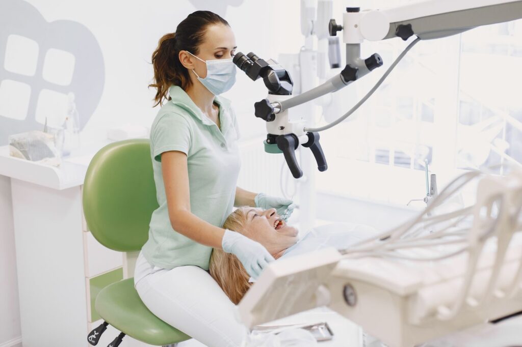
    About this guide. Written for patients of Southeast Endodontics, PC, Charleston, SC. Night guards are usually fitted by your general dentist; we see the consequences when a grinding-related crack reaches the pulp.

  • Why Does My Tooth Hurt When I Bite Down?

    Why Does My Tooth Hurt When I Bite Down?

    Pain on biting is one of the more useful symptoms you can bring to a dentist, because the exact way it behaves narrows the diagnosis faster than almost anything else.

    Whether it hurts pressing down or letting go, whether it is one tooth or several, and whether it started after dental work all point in different directions. Here is how to read it.

    Key takeaways

    • Pain on releasing a bite suggests a crack; pain on pressing down suggests the ligament.
    • Bite pain starting days after a filling usually means the restoration is fractionally high.
    • An inflamed ligament makes a tooth tender with nothing visible on an X-ray.
    • Clenching produces the same symptom with no decay, crack or infection at all.

    What the timing tells you

    Pay attention to the exact moment it hurts. Patients often describe this precisely once asked, and it is genuinely diagnostic.

    What each pattern of bite pain usually means
    When it hurtsMost likely causeWhat usually fixes it
    On releasing the biteA crack flexing open and snapping shutCrown; root canal first if the pulp is involved
    On pressing down, dull and constantInflamed ligament at the root tipRoot canal treatment or retreatment
    Only since a recent fillingRestoration fractionally too highA minute of bite adjustment
    On hard food onlyEarly crack, or a worn cuspAssessment; often a crown
    Several teeth, worse in the morningClenching or grindingA night guard and stress management
    With swelling or a gum bumpInfection at the root tipSame-day drainage and treatment

    Swipe the table sideways to see every column.

    Pain on release: the classic crack

    This is the pattern worth learning. Chewing something firm produces a jolt not while you bite but as you let go — the crack closes and momentarily pinches the pulp.

    Pain on releasing the bite, rather than on pressing down, suggests a crack
    Firm foods produce it; soft foods rarely do.

    It is often intermittent, food-dependent and hard to localise, which is why people put up with it for months. Cracks propagate under load, so a tooth that needs only a crown now may need considerably more later — which cracked teeth can be saved covers the five types and their prognosis.

    Pain on pressure: the ligament

    Each tooth is suspended in its socket by the periodontal ligament. When infection or inflammation from inside the tooth reaches the root tip, that ligament becomes inflamed and exquisitely pressure-sensitive.

    An inflamed ligament makes even light contact painful
    An inflamed ligament makes even light contact painful, with little to see on an early radiograph.

    The result is a dull, constant tenderness — the tooth may feel raised, as though it is hitting first. This is the pattern most often associated with a dying or dead pulp, and it can appear before anything shows on a radiograph. It also occurs after root canal treatment as normal healing, which our recovery guide covers.

    Pain that started after dental work

    This is the easiest cause to fix and the one people most often endure unnecessarily. A new filling or crown that sits slightly proud means that tooth contacts first, every time.

    A filling a fraction too high takes the whole force of your bite
    A filling a fraction of a millimetre high takes the full force of your bite before any other tooth.

    The ligament becomes bruised from the constant overload, and the tooth feels sore to bite on. Adjustment takes a minute or two, and if the pain resolves within days afterwards, that confirms the diagnosis. It is worth ringing whoever placed the restoration rather than waiting it out.

    When it is clenching, not the tooth

    If several teeth are tender, the pain is worse in the morning, and your jaw muscles ache, the cause may not be in any tooth at all.

    Clenching overloads the ligament without any decay being present
    Clenching overloads the ligament with no decay, crack or infection involved.

    Sustained clenching overloads the ligaments of multiple teeth at once. Nothing shows on a radiograph because nothing is wrong with the teeth — see how grinding damages teeth for what to do about it.

    How it is diagnosed

    The central test is simple: you bite on a small plastic wedge placed on one cusp at a time. A crack usually produces pain on one cusp specifically, and on release rather than pressure.

    Testing each cusp separately localises the problem
    Testing each cusp separately localises which part of which tooth is involved.

    Percussion testing — light tapping — identifies ligament inflammation, and cold testing establishes whether the pulp is alive. Together these separate the causes above in a few minutes. Our guide to what temperature sensitivity means covers the pulp side.

    What happens at the appointment

    Diagnosis of bite pain is mostly clinical rather than radiographic, which surprises people expecting an X-ray to settle it.

    The tests used, and what each one shows
    TestWhat it involvesWhat it tells us
    Bite stick, cusp by cuspBiting a small plastic wedge on each cusp in turnLocalises a crack to one cusp, and whether pain is on release
    PercussionLight tapping on the toothInflammation of the ligament at the root tip
    PalpationPressing the gum over the rootInfection spreading into surrounding bone
    Cold testingCold applied briefly to the toothWhether the pulp is alive, and how it reacts
    TransilluminationLight shone through the toothA crack interrupts the glow
    RadiographStandard or 3D imagingBone changes; most cracks do not show at all

    Swipe the table sideways to see every column.

    That last row is worth absorbing. A normal-looking X-ray does not rule out a crack or early ligament inflammation, and being told the film looks fine is not the same as being told nothing is wrong.

    When to be seen quickly

    Bite pain on its own justifies an appointment within days rather than hours. Cracks worsen under load and an inflamed ligament rarely settles unaided.

    Most causes are straightforward once the right one is identified
    Most causes of bite pain are straightforward once the right one is identified.

    Move faster if there is facial swelling, fever, a bump on the gum, or pain that wakes you — those indicate infection reaching the bone, covered in our abscess guide and the emergency symptom list.

    If a tooth has been sore to bite on for more than a week or two, an assessment is worth having — see what a consultation involves. The Merck Manual’s entry on pulpitis covers the underlying condition clinically.

    Frequently asked questions

    My tooth only hurts when I let go of a bite. What does that mean?

    That specific pattern points to a crack. Biting wedges the crack open; releasing lets it snap shut and pinches the pulp inside for a fraction of a second. It is the most characteristic symptom in dentistry.

    I had a filling last week and now it hurts to bite. Is that normal?

    Briefly, yes — but not for long. If it persists beyond a few days, the filling is probably fractionally high and taking your bite force first. Adjusting it takes minutes and the relief is usually immediate.

    Can I have pain on biting with no decay and no crack?

    Yes. An inflamed ligament around the root tip does exactly this, and so does clenching. Both make a tooth tender to pressure without anything visible on an X-ray at first.

    How urgent is it?

    Depends on what comes with it. Pain on biting alone warrants an appointment within days. Add swelling, fever, or pain that wakes you and it becomes same-day.

    Have Questions or Ready to Schedule?

    Our team is here to help — call, email, or request an appointment online.

    About this guide. Written for patients of Southeast Endodontics, PC, Charleston, SC. The patterns above are guides rather than a diagnosis — several causes can coexist in the same tooth.

  • Apicoectomy: What Root-End Surgery Actually Involves

    Apicoectomy: What Root-End Surgery Actually Involves

    When a root canal has been done well and the tooth still will not settle, the problem is usually at the very tip of the root — the one part that cannot always be reached from inside the tooth.

    An apicoectomy solves that by approaching from the outside instead. Here is what the procedure involves, what recovery is actually like, and when it is the right answer rather than retreatment or extraction.

    Key takeaways

    • The root tip and surrounding infected tissue are removed through the gum, and the root end is sealed.
    • It takes 30–90 minutes and is done under local anaesthetic.
    • Microsurgical technique reports around 90% success or better — far above older published figures.
    • It is a second-line option: non-surgical retreatment is normally tried first.

    Why the root tip is the problem

    A root canal cleans and seals the canal from the crown downwards. At the very end of the root, the canal frequently stops being a single tube and becomes a fine delta of branches too narrow for any instrument to enter.

    Surgery is considered when re-entering the tooth is not realistic
    Surgery is considered when re-entering the tooth is not realistic, or has already been tried.

    Bacteria surviving in that mesh keep the surrounding bone inflamed. No amount of cleaning from inside reaches them, which is why a technically excellent root canal can still leave a tooth that aches on biting or shows a persistent shadow on a radiograph.

    When it is the right choice

    Apicoectomy is not the first response to a failing root canal. Non-surgical retreatment is generally tried first, because it addresses the whole canal rather than just the end of it.

    When surgery is preferred over retreatment
    SituationWhy retreatment is unsuitable
    A well-fitting post and crownRemoving them risks fracturing the root or destroying a sound restoration
    A separated instrument blocking the canalThe blockage cannot be bypassed from inside
    Retreatment already attempted and failedThe canal has been cleaned as far as it can be
    A cyst or lesion needing biopsyTissue can only be removed and examined surgically
    A perforation low in the rootRepair from outside is more predictable

    Swipe the table sideways to see every column.

    What actually happens

    1. Anaesthetic. The area is numbed thoroughly, including the surrounding bone.
    2. Access. A small incision is made in the gum beside the tooth and the tissue lifted to expose the bone over the root tip.
    3. Removal. A few millimetres of the root tip are removed, along with the infected tissue around it.
    4. Sealing. The cut end of the root is prepared and sealed with a biocompatible filling material.
    5. Closure. The gum is repositioned and closed with fine sutures, usually removed after a week.
    A small, precise procedure rather than a major operation
    A precise, small-field procedure rather than a major operation.

    Magnification is the single biggest change in this procedure over the last twenty years. Working under a microscope allows a much smaller opening, a cleaner cut and a far better seal — see the equipment we use.

    Microsurgical technique is what lifted success rates above 90%
    Microsurgical technique is what lifted reported success rates above 90%.

    Recovery, honestly

    Expect soreness and swelling. It peaks at about 48 hours and then improves daily, and a cold compress in the first day genuinely helps.

    Swelling peaks around 48 hours and settles within a few days
    Swelling peaks around 48 hours, then settles.
    Apicoectomy recovery timeline
    WhenWhat to expectWhat to do
    Day of surgeryNumbness, then soreness as it wears offCold compress, anti-inflammatories, rest
    Day 1–2Swelling peaks; possible bruisingSoft food, no vigorous rinsing or brushing at the site
    Day 3–7Steady improvement; sutures removed around day 7Return to normal activity; gentle cleaning
    Week 2–4Soft tissue fully healedNormal brushing and diet
    Month 3–6Bone fills in at the root tipFollow-up radiograph to confirm healing

    Swipe the table sideways to see every column.

    Soft food and gentle rinsing for the first few days
    Soft food and gentle cleaning for the first few days; most people are back to normal activity the next day.

    Most patients return to work the following day. What takes longer is invisible: the bone defect left by the infection fills in over three to six months, which is why we review with a radiograph rather than judging by symptoms.

    What the success rate really means

    You will find figures ranging from 60% to 97% for this procedure, and the spread is not noise — it is chronology.

    Older studies describe surgery done without magnification, with materials long superseded. Contemporary microsurgical series report around 90% or better, with roughly 97% still successful at five years and more than 75% at ten to thirteen years. If you are reading an alarming number, check when the study was published.

    What it costs

    An apicoectomy typically runs somewhere between a molar root canal and an extraction with an implant. It is billed as a surgical endodontic procedure, which most plans cover at the same percentage as other endodontic work — though it draws on the same annual maximum.

    Ask for a written estimate with your benefits checked before treatment. Our cost guide explains how deductibles and annual maximums interact, and why timing across two plan years sometimes helps.

    The risks worth knowing about

    It is minor surgery, and minor surgery is not no surgery. Bruising and swelling are expected rather than complications.

    The specific risks depend on where the tooth sits. Upper back teeth are close to the maxillary sinus, and lower back teeth to the nerve supplying the lip and chin, so we assess both on 3D imaging before operating rather than discovering the relationship during the procedure. Numbness of the lip is uncommon and usually temporary where it occurs.

    The other honest caveat is that not every case is suitable. A tooth with a vertical root fracture, or too little bone support remaining, will not be improved by surgery — and we would tell you that rather than attempt it.

    Weighing it against extraction

    The honest comparison is not surgery versus doing nothing, but surgery versus removing the tooth and replacing it.

    Bone at the root tip rebuilds over three to six months
    The tooth keeps its own root and ligament, which nothing replaces.

    An apicoectomy keeps your own root, ligament and bone, and costs considerably less than extraction plus an implant. It is also reversible in the sense that extraction remains available afterwards — the reverse is not true. The alternatives compared on ten-year cost sets out the numbers.

    Our apicoectomy procedure page covers the clinical detail, and endodontic surgery the wider category. The published analysis of outcomes after non-surgical treatment fails sets surgery in context against the alternatives.

    Frequently asked questions

    Is an apicoectomy painful?

    During, no more than a root canal — the area is fully anaesthetised. Afterwards expect soreness and swelling for a few days, managed with anti-inflammatories and a cold compress. Most people describe it as comparable to having a tooth out, without losing the tooth.

    How long does it take?

    Between 30 and 90 minutes, depending on which tooth and how accessible the root tip is. Front teeth are usually at the shorter end; lower molars at the longer end.

    What is the success rate?

    Reported success with modern microsurgical technique is around 90% or better, with roughly 97% still doing well at five years in some series. Older figures quoting 60–70% predate the microscope and current materials.

    Can it be repeated if it fails?

    Sometimes, but a second surgery on the same root has a lower success rate. If an apicoectomy fails, extraction and replacement is usually the more sensible next step than a repeat attempt.

    Have Questions or Ready to Schedule?

    Our team is here to help — call, email, or request an appointment online.

    About this guide. Written for patients of Southeast Endodontics, PC, Charleston, SC. Success figures are published averages; whether surgery suits your tooth depends on its anatomy, restoration and treatment history.

  • Loose or Chipped Tooth After an Accident: What to Do

    Loose or Chipped Tooth After an Accident: What to Do

    A knocked-out tooth is obviously an emergency. A tooth that is merely loose, pushed slightly out of line, or chipped is the injury people talk themselves out of getting checked — and it is the one that quietly goes wrong months later.

    Seek care today if a tooth is visibly displaced, loose enough to move when you touch it, or the chip exposes a pink or red spot in the middle of the tooth.

    Go to hospital first if there was loss of consciousness, a suspected jaw fracture, or an injury needing medical attention. The tooth can be dealt with afterwards.

    Key takeaways

    • A loose or displaced tooth should be repositioned and splinted, ideally the same day.
    • Splinting is short and flexible — about two weeks for subluxation, four for luxation.
    • A chip exposing yellow dentine or a pink spot needs treating within hours, not days.
    • The pulp can die quietly months later, which is why follow-up runs for a year.

    The injuries, and how each is handled

    Dental injuries short of avulsion, and what each needs
    InjuryWhat you noticeUsual treatment
    ConcussionTender to touch, not looseMonitoring; soft diet
    SubluxationLoose but not moved out of positionFlexible splint about 2 weeks
    Extrusive luxationTooth looks longer, feels high on bitingRepositioned, splinted around 4 weeks
    Lateral luxationPushed sideways, often locked in placeRepositioned, splinted around 4 weeks
    Intrusive luxationDriven up into the socket, looks shorterDepends on root maturity; often needs specialist care
    Enamel chipRough edge, no colour changeSmoothing or bonding, not urgent
    Chip exposing dentine or pulpYellow area, or a pink or red spotSame-day protection of the pulp

    Swipe the table sideways to see every column.

    Why a loose tooth matters even without pain

    The periodontal ligament suspends each tooth in its socket. An impact tears some of those fibres, and the tooth loosens.

    A flexible splint holds the tooth while the ligament reattaches
    A flexible splint holds the tooth to its neighbours while the ligament reattaches.

    Left unsupported, it moves during healing and the ligament reattaches poorly. Worse, the blood vessels entering the root tip may have been stretched or severed — which the tooth will not tell you about for weeks.

    A splint is a thin wire or composite bonded to the injured tooth and the sound teeth either side. Deliberately flexible: some physiological movement encourages normal ligament healing, whereas rigid fixation for too long risks the tooth fusing to bone.

    Reading a chipped tooth

    Size is a poor guide. What matters is which layer is exposed.

    A chip confined to enamel is cosmetic; a chip exposing dentine is not
    How deep the chip goes matters far more than how big it looks.
    • Enamel only — a rough edge, uniform colour. Cosmetic; bond or smooth it at your convenience.
    • Into dentine — a distinctly yellower area. Sensitive to air and cold, and needs covering within a day or two because dentine tubules lead to the pulp.
    • Pulp exposed — a pink or red dot, possibly bleeding. Same-day treatment. In a young tooth, prompt care can keep the pulp alive.

    Keep any fragment in milk and bring it — it can often be bonded back. If the tooth was also loosened, both problems need addressing at the same visit.

    Why the pulp can fail months later

    This is the part patients find hardest to accept when the tooth feels fine. Trauma stretches the vessels entering the root tip; they may recover, or thrombose and cut off the blood supply.

    The pulp can die quietly weeks or months after the accident
    The pulp can survive the injury, or die quietly weeks later. Testing is the only way to know.

    If the pulp dies, it does so without symptoms in many cases. The first sign is often the tooth darkening — see why a tooth turns dark — or an abscess appearing at the root tip long after the accident is forgotten.

    What to do in the first hour

    Do not try to push a displaced tooth back into position yourself. Unlike a knocked-out tooth, where immediate replanting genuinely helps, a luxated tooth is still attached and forcing it risks further damage to the ligament and the bone around it.

    Instead: bite gently on clean gauze to steady it, use a cold compress on the outside of the face for the swelling, take ibuprofen if it suits you, and eat nothing that needs chewing until you have been seen.

    If a fragment broke off, keep it moist in milk and bring it. Reattaching the patient’s own fragment gives a better colour and translucency match than any composite, and it is often still possible hours later.

    Photograph the injury before anything is treated if you can. It is genuinely useful for insurance, and for comparison at the follow-up appointments.

    The follow-up schedule, and why it is that long

    What each follow-up is checking for
    WhenWhat we checkWhy then
    2 weeksSplint, pulp response, healingSplint removal for subluxation
    4 weeksPulp testing, mobilitySplint removal for luxation injuries
    8 weeksPulp vitality, early resorptionNecrosis often first detectable here
    6 monthsVitality, root development in childrenLate necrosis and resorption
    1 yearRadiographic reviewConfirms stable long-term healing

    Swipe the table sideways to see every column.

    Reviews at 2 and 4 weeks, then 8 weeks, 6 months and a year
    Two weeks, four weeks, eight weeks, six months, one year.

    If the pulp does die, root canal treatment resolves it, and in an immature tooth the approach differs — see endodontics in children. Our traumatic injuries page covers the wider clinical picture.

    What recovery usually looks like

    Expect the tooth to feel tender to bite on for a week or two after splinting, and slightly loose for a while after the splint comes off. Both settle.

    Eat soft food while splinted, keep the area meticulously clean, and avoid contact sport until you are discharged. Most displaced teeth recover fully when they are repositioned early and reviewed properly.

    Prevention, since most of these are preventable

    A properly fitted mouthguard prevents the majority of sports-related dental injuries, including the displacements described here, and costs a fraction of treating one.

    Bicycle falls and playground impacts account for most of these injuries
    Bicycle falls, sport and playground impacts account for most of what we see.

    Cleveland Clinic’s overview of tooth luxation is a useful independent reference on the injury types above.

    Most displaced teeth survive if they are repositioned and monitored
    Repositioned early and monitored properly, most displaced teeth survive.

    Frequently asked questions

    My tooth is loose after a knock but does not hurt. Do I still need to be seen?

    Yes, and soon. Mobility means the ligament holding the tooth has been damaged, and a tooth that stabilises on its own can still lose its pulp quietly over the following months. Splinting early gives it the best chance.

    How long does a splint stay on?

    It depends on the injury. Around two weeks for a subluxation, roughly four weeks for extrusive or lateral luxation. Longer is not better — prolonged rigid splinting increases the risk of the tooth fusing to the bone.

    I chipped a tooth and it looks fine. Can I leave it?

    If the chip is confined to enamel, it is cosmetic and not urgent. If you can see yellow dentine, or a pink or red spot in the middle, the pulp is exposed or close to it and that needs treating within hours.

    Why do I need follow-ups for a year?

    Because pulp necrosis after trauma frequently appears late. Reviews at two weeks, four weeks, eight weeks, six months and a year catch a dying pulp before it becomes an abscess.

    Have Questions or Ready to Schedule?

    Our team is here to help — call, email, or request an appointment online.

    About this guide. Written for patients of Southeast Endodontics, PC, Charleston, SC, following International Association of Dental Traumatology guidance. If an injury happened today, call rather than finish reading.

  • Root Canals in Children: Baby Teeth and Immature Roots

    Root Canals in Children: Baby Teeth and Immature Roots

    Endodontic treatment in children is not simply the adult procedure scaled down. Their teeth are still developing, and the treatment has to work with that rather than against it.

    Here is what changes with a child’s tooth, why we often treat baby teeth people expect us to pull, and how an unfinished root is handled.

    Key takeaways

    • Baby teeth hold space for the adult teeth behind them — losing one early causes crowding.
    • A pulpotomy treats only the pulp in the crown and leaves the roots alone.
    • An immature permanent tooth has an open root tip that cannot be sealed conventionally.
    • A knocked-out baby tooth should never be replanted.

    Why baby teeth are worth treating

    The instinct to pull a decayed baby tooth is understandable and usually wrong. Primary molars stay in place until around age eleven or twelve, and they are holding the space the permanent premolar will need.

    Remove one early and the adjacent teeth drift into the gap. The permanent tooth then erupts crowded, rotated, or not at all — converting a treatable tooth into years of orthodontics.

    Pulpotomy: the usual treatment for a baby tooth

    Where decay has reached the pulp of a primary tooth but the roots are still healthy, a pulpotomy removes the inflamed tissue in the crown, places a medicated dressing over what remains, and seals it — usually with a stainless steel crown.

    A pulpotomy treats the crown portion of the pulp and leaves the rest
    Only the pulp inside the crown is removed; the root portion stays.

    It is quicker and less invasive than full root canal treatment, and it leaves the root portion of the pulp intact so the tooth resorbs and exfoliates naturally when the time comes.

    Treatment options for children’s teeth, by situation
    SituationUsual treatmentWhat it preserves
    Deep decay, pulp not yet exposedIndirect pulp capThe whole pulp
    Small pulp exposure, healthy pulpDirect pulp cap or partial pulpotomyMost of the pulp
    Inflamed crown pulp, healthy rootsPulpotomy plus crownRoot pulp; natural exfoliation
    Whole pulp necrotic, primary toothPulpectomy with resorbable fillingThe tooth and its space
    Tooth unrestorableExtraction plus space maintainerThe space, not the tooth

    Swipe the table sideways to see every column.

    When the root has not finished forming

    Permanent teeth erupt before their roots are complete. It takes two to three more years for the tip to close, and during that window a tooth is genuinely awkward to treat.

    An immature root has an open tip that has not finished forming
    An immature root has a wide open tip and thin walls.

    The problem is mechanical: there is no narrowing at the root tip to fill against, so conventional sealing is not possible. Thin root walls also make the tooth prone to fracture.

    Approaches to an immature permanent tooth
    ApproachWhen it is usedWhat it achieves
    ApexogenesisPulp still vitalRoot keeps developing normally
    PulpotomyInflamed but vital pulpAround 86% success in immature teeth
    Regenerative endodonticsNecrotic pulp, open apexBest reported root maturation of the three
    ApexificationNecrotic pulp, regeneration unsuitableBuilds a barrier so the canal can be sealed

    Swipe the table sideways to see every column.

    Apexification builds a barrier so the canal can be sealed
    Apexification creates a hard barrier at the tip so the canal can finally be sealed.

    Where the pulp is still alive, keeping it alive is always the goal — a vital pulp continues to lay down dentine and thicken the root walls, which no artificial material replicates.

    Why children’s teeth behave differently

    Two features of a young tooth change the clinical picture entirely, and both work in the child’s favour.

    The pulp chamber is proportionally much larger, which means decay reaches it sooner but also that the pulp has a far richer blood supply and a genuine capacity to heal. Treatments that would be optimistic in an adult are realistic in a child.

    The roots are also still forming, or in the case of baby teeth already resorbing on a schedule. Treatment has to account for where that tooth is in its life cycle rather than treating it as a small adult tooth.

    Trauma is the commonest reason we see children

    Sport, playgrounds and bicycles account for most childhood dental injuries, and upper front teeth take the brunt. Those teeth are often the ones with immature roots, which is why children’s trauma frequently lands with an endodontist.

    Trauma to a developing front tooth is the commonest reason we see children
    Upper front teeth take the impact in most childhood accidents.

    If a permanent tooth is knocked out, the first thirty minutes matter enormously — our avulsed tooth guide covers the protocol. For displaced or chipped teeth, see what to do after an impact. Baby teeth are the exception: never replant one.

    A mouthguard prevents most of these injuries, and our tips for protecting natural teeth cover choosing one.

    What to watch for at home

    Children under-report dental pain, and they adapt to it. A child who has quietly stopped using one side to chew is telling you something even if they say nothing hurts.

    Worth an appointment: a tooth that has changed colour after a knock, a persistent pimple on the gum, swelling of the face or gum, waking at night, or avoiding cold food. Any of those in a child means the same as in an adult.

    After any facial impact, keep watching for months rather than days. A front tooth that darkens six months after a playground fall is the commonest late presentation we see, and the accident is usually long forgotten by then.

    What the appointment is like

    A parent or guardian accompanies every patient under 18, and stays through the consultation. We explain what we are doing in terms the child can follow, because surprise is what frightens children rather than the procedure itself.

    A parent or guardian stays with any patient under 18
    A parent or guardian stays with any patient under 18 throughout.

    Where anxiety is significant, nitrous oxide works well for children and wears off within minutes. Our guide to managing dental anxiety covers the wider options. The pulp-preserving approach used in children is now used selectively in adults too — see vital pulp therapy in mature teeth.

    Keeping a baby tooth until it is ready to go protects the adult tooth behind it
    Keeping a baby tooth until it is ready to go protects the adult tooth behind it.

    If your child has toothache, a discoloured front tooth after a knock, or an injury you are unsure about, it is worth an assessment — contact the practice. The American Academy of Pediatric Dentistry publishes its clinical guideline on pulp therapy for primary and immature permanent teeth.

    Frequently asked questions

    Why treat a baby tooth that is going to fall out anyway?

    Because it is holding space. A primary molar lost early lets the teeth behind it drift forward, and the permanent tooth underneath then has nowhere to erupt. That is an orthodontic problem created to avoid a filling.

    Is a pulpotomy the same as a root canal?

    No. A pulpotomy removes only the inflamed pulp in the crown and leaves the healthy pulp in the roots. A root canal removes all of it. On children’s teeth the partial approach is usually enough and far less invasive.

    What happens if a child knocks out a baby tooth?

    Do not put it back. Replanting a primary tooth can damage the permanent tooth developing above it. See a dentist to check nothing else is injured, but leave the tooth out.

    Can a child have a root canal on a permanent tooth?

    Yes, but if the root is not fully formed it needs a different approach. An open root tip cannot be sealed conventionally, so treatment aims to let the root finish developing or to build a barrier at the tip.

    Have Questions or Ready to Schedule?

    Our team is here to help — call, email, or request an appointment online.
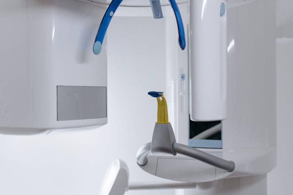
    About this guide. Written for parents by Southeast Endodontics, PC, Charleston, SC. Which treatment suits a child’s tooth depends on their age, the stage of root development and the state of the pulp.

  • Endodontist or General Dentist: Who Should Treat Your Tooth?

    Endodontist or General Dentist: Who Should Treat Your Tooth?

    Your dentist says you need a root canal and offers to do it. Should you say yes, or ask to see a specialist? It is a fair question, and the honest answer depends almost entirely on which tooth it is.

    Here is what actually differs between the two, what the outcome data shows, and the cases where it genuinely matters.

    Key takeaways

    • An endodontist is a dentist plus two to three years of residency limited to this procedure.
    • Reported success is roughly 90–95% for specialists against 85–90% for general practice — though specialists take the harder cases.
    • For a straightforward front tooth, your own dentist is very likely the right choice.
    • Molars, retreatments, unclear diagnoses and “we could not get you numb” are specialist territory.

    What the training difference actually is

    Both are dentists. The difference is what happened afterwards: a residency limited entirely to endodontics, covering diagnosis of orofacial pain, complex canal anatomy, retreatment and surgical endodontics.

    Two to three years of additional training beyond dental school
    Two to three years of full-time residency, after dental school, doing nothing else.

    Volume is the part patients underestimate. A dentist may graduate having completed a handful of root canals; an endodontist typically finishes residency having done several hundred, and then does little else for the rest of their career.

    What the outcome data shows

    Reported root canal outcomes, specialist vs. general practice
    MeasureEndodontistGeneral dentistCaveat
    Reported success rate~90–95%~85–90%Specialists treat harder cases, which understates the gap
    Cases completed in trainingSeveral hundredOften fewer than tenVolume compounds over a career
    Microscope used routinelyStandardLess commonMissed canals are a leading cause of failure
    3D imaging on siteUsualVariesMatters most for molars and retreatment
    Typical fee difference10–20% higherBaselineOften reimbursed at the same rate

    Swipe the table sideways to see every column.

    Read that table carefully in one respect: the success gap is measured across different case mixes. Specialists receive the curved, calcified and previously failed teeth, which drags their average down relative to a like-for-like comparison.

    When your own dentist is the right answer

    Plenty of root canals are genuinely routine, and there is no advantage in paying more for one. Straightforward cases share a profile: a front tooth or premolar, a single canal visible along its whole length on the radiograph, a clear diagnosis, and no history of difficulty getting numb.

    A referral is a judgement about the case, not about your dentist
    A referral is a judgement about the case, not a criticism of your dentist.

    Many general dentists do this work very well and enjoy it. If yours is confident about a case that fits that description, that confidence is usually well founded.

    When it is worth seeing a specialist

    Situations where a specialist referral is worth requesting
    SituationWhy it matters
    Molar, especially a lower first molarThree to four curved canals; the extra canal is easy to miss
    Previous root canal on the toothRetreatment needs the old filling removed before anything else
    You could not get numb last timeHot pulps need supplementary anaesthetic technique
    Nobody is sure which tooth it isReferred pain; treating the wrong tooth is the costly error
    A crack is suspectedDiagnosis relies on magnification and testing, not imaging
    The tooth is calcified on the radiographCanals may be difficult or impossible to locate without a microscope

    Swipe the table sideways to see every column.

    Molars, retreatments and unclear diagnoses are where the difference shows
    Molars, retreatments and unclear diagnoses are where the difference shows up.

    The equipment argument is not marketing. Our page on the technology used here covers it, and why root canals fail explains how often the cause turns out to be anatomy nobody could see.

    Magnification and 3D imaging are standard equipment in a specialist practice
    A missed canal is a leading cause of failure, and magnification is how they are found.

    What a specialist practice does differently

    The equipment matters less than how routinely it is used. A microscope that comes out for difficult cases is not the same as one used on every tooth from the first minute.

    Scheduling differs too. A practice doing only endodontics books the time a molar actually needs rather than fitting it between check-ups, which is why specialist appointments are usually longer and less often split across visits.

    Anaesthesia is the third difference, and the one patients notice. Supplementary techniques for teeth that will not go numb — intraosseous and intraligamentary injections among them — are routine in a specialist practice and less commonly needed in general practice.

    What it costs, honestly

    Expect to pay 10–20% more, commonly $200–$400 extra on a molar. That is a genuine cost and worth weighing.

    Most plans reimburse specialist and general fees at the same rate
    The fee difference is real; the out-of-pocket difference is often smaller.

    Two things reduce it. Most insurance plans reimburse endodontic treatment at the same percentage regardless of who provides it, and a case that has to be redone costs far more than the original difference — the full cost breakdown covers both.

    What happens to the tooth afterwards

    One practical point is worth knowing before you choose, because it surprises people who expect a specialist to handle everything.

    We complete the root canal and place a temporary filling. Your own dentist then places the permanent restoration, which on a back tooth means a crown. So seeing a specialist does not replace your dentist — it adds one appointment in the middle of a course of treatment that returns to them.

    That split is also why the referral note back matters. You should expect a written report and post-treatment radiographs to reach your dentist, so the restorative work can start without repeating anything.

    How to raise it with your dentist

    Ask directly and without awkwardness: how many canals does this tooth have, how confident are you about this one, and would you refer it if it were your own tooth? Those are ordinary clinical questions. So is asking someone else — how a second opinion works sets out where to take which question.

    The gap in reported success is real but smaller than marketing suggests
    The right question is not who is better, but which case this is.

    Dentists refer routinely, and the good ones welcome being asked. If you would rather come to us first, you can — see what a first visit involves and meet Dr. Long and Dr. O’Neal. For colleagues, our guide to referral criteria covers the same ground clinically.

    The American Association of Endodontists sets out its own account of why and when it is worth seeing an endodontist, which is worth reading alongside this page rather than instead of it.

    Frequently asked questions

    Is an endodontist a real dentist?

    Yes. An endodontist completes dental school first, then two to three further years of specialty residency limited to endodontics. It is the same qualification plus a specialty on top, not a different profession.

    Does it cost more to see a specialist?

    Usually 10–20% more per procedure — often $200–$400 on a molar. Most plans reimburse specialist and general fees at the same rate, so the out-of-pocket difference is frequently smaller than the fee difference suggests.

    Do I need a referral?

    Not with us. Many patients arrive by referral from their general dentist, but you can book directly. Either way we send a treatment report back to your dentist so the restorative work can follow.

    Will my dentist be offended if I ask to be referred?

    No, and it is a reasonable question to ask. Referring is a normal part of practice, and most dentists refer the cases they judge to be outside their comfortable range as a matter of routine.

    Have Questions or Ready to Schedule?

    Our team is here to help — call, email, or request an appointment online.
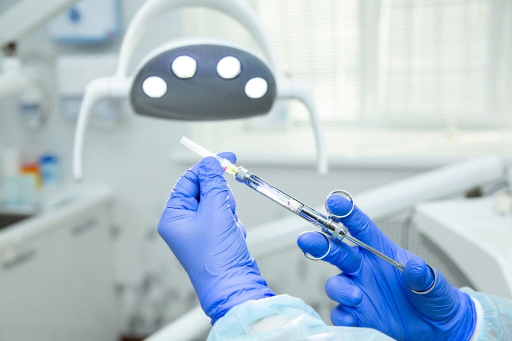
    About this guide. Written by Southeast Endodontics, PC, Charleston, SC. We are a specialist practice, so read the above with that in mind — which is exactly why the section on when your own dentist is the right choice is included.

  • Sinus Pain or Toothache? How to Tell the Difference

    Sinus Pain or Toothache? How to Tell the Difference

    Upper back teeth that ache during a heavy cold are one of the most commonly misdiagnosed complaints in dentistry — in both directions. People have root canals they did not need, and sinus treatment that was never going to work.

    The two problems sit millimetres apart anatomically. Here is how to tell them apart, and when it turns out to be both.

    Key takeaways

    • Upper molar roots sit directly beneath the maxillary sinus, sometimes with paper-thin bone between them.
    • Sinus pain typically affects several upper teeth; a tooth problem is nearly always one tooth.
    • Pain that worsens when you bend forward points to sinus. Toothache does not care about posture.
    • More than 40% of maxillary sinus infections actually start in a tooth.

    Why the confusion happens

    The maxillary sinuses are air spaces behind your cheekbones. The roots of your upper premolars and molars push up into the floor of those spaces, occasionally protruding through it entirely.

    Upper molar roots sit directly beneath the maxillary sinus floor
    Upper molar roots project into the sinus floor, sometimes with almost no bone between them.

    Both are supplied by branches of the same nerve, and the brain is poor at distinguishing where pain within a shared nerve territory originates. That is the whole problem in one sentence.

    Six ways to tell them apart

    Sinus pain vs. tooth pain: the distinguishing features
    FeaturePoints to sinusPoints to the tooth
    How many teethSeveral upper teeth at onceOne tooth, usually identifiable
    CharacterDull, heavy, pressure-likeSharp, throbbing, or jabbing
    Bending forwardNoticeably worseNo change
    Hot and coldLittle effectOften the main trigger
    Biting on itUsually fineFrequently painful
    Other symptomsCongestion, discharge, reduced smell, facial pressureNone of those
    Both sides?Often bilateralAlmost always one side

    Swipe the table sideways to see every column.

    Nasal congestion alongside the pain points away from the tooth
    Nasal congestion alongside the ache is the single most useful clue.

    The bending-forward test is worth doing deliberately. Lean down as if tying a shoelace and hold it for a few seconds. Sinus pressure builds noticeably; a tooth problem does not change.

    Sinus pain typically worsens when you bend forward; toothache does not care
    Position-dependent pain is a sinus signature.

    The overlap nobody mentions

    Real life is less tidy than the table above, and two situations blur it further.

    The first is a genuinely coincidental pair. Sinusitis is common and toothache is common, and having both at once is not rare. If sinus treatment resolves the congestion but one tooth still aches, that remaining tooth deserves its own assessment rather than another course of decongestants.

    The second is recent dentistry. An upper molar extracted or treated close to the sinus floor can leave a small communication between mouth and sinus, and sinus symptoms that begin within days of such a procedure should go back to whoever performed it.

    In both cases the useful instinct is the same: treat the finding you can actually demonstrate, and re-examine rather than assume when symptoms only partly resolve.

    When it is genuinely the tooth

    Certain features effectively rule sinus out. If cold makes it worse and the pain lingers well after the cold is gone, that is pulpal — our guide to what hot and cold sensitivity means covers how to read it.

    Cold testing separates a live pulp from a sinus problem in seconds
    Cold testing separates a live pulp from a sinus problem in seconds.

    Sharp pain on releasing a bite suggests a crack. Pain that wakes you at night, or swelling of any kind, points firmly at the tooth and needs seeing quickly — see which symptoms are urgent.

    When the tooth caused the sinusitis

    This is the case most often missed. An infected upper molar can discharge directly into the sinus above it, producing genuine sinusitis with a dental cause — maxillary sinusitis of dental origin.

    More than 40% of maxillary sinus infections start in a tooth
    A dental origin is easy to miss when the presentation looks purely sinus.

    The pattern to watch for is one-sided sinusitis that keeps returning despite antibiotics or nasal treatment, often with an unpleasant taste or smell. Treating the sinus alone never resolves it, because the source is a tooth.

    Which clinician to start with
    Your situationStart withWhy
    Cold or allergy symptoms, several upper teeth acheYour doctorLikely sinusitis; teeth are referred pain
    One tooth, reacts to hot or coldA dentist or endodontistClassic pulpal presentation
    One-sided sinusitis that keeps returningA dentist firstSuspect a dental origin
    Recent dental work on that side, now sinus symptomsThe treating dentistCould be related to the procedure
    Facial swelling, fever, or the eye area involvedEmergency care todaySpreading infection, whatever the source

    Swipe the table sideways to see every column.

    Why guessing is expensive

    Both errors happen, and both cost more than the assessment would have.

    Treating a healthy tooth for sinus pain is irreversible. Root canal treatment on a tooth whose pulp was never the problem removes healthy tissue and leaves the actual pain untouched, which is how people end up having a second and third tooth treated in search of relief.

    The reverse wastes time rather than tooth structure. Weeks of decongestants and antibiotics for what is actually an infected molar allow the infection to progress, and the tooth that could have been treated straightforwardly becomes an abscess with facial swelling.

    Neither is a criticism of anyone. The presentations genuinely overlap, which is precisely why testing rather than reasoning settles it.

    How the diagnosis is actually made

    Testing settles it quickly. Cold testing shows whether the pulp is alive and how it responds; percussion testing shows whether inflammation has reached the ligament around the root.

    Treating the wrong one wastes weeks; the tests take minutes
    The tests take minutes; treating the wrong problem takes weeks.

    A live, normally responding pulp on every upper tooth in the area effectively rules out a dental cause. Where the picture is unclear, 3D imaging shows the relationship between root tips and the sinus floor directly — see the imaging we use.

    If you are unsure, an assessment is worth having before committing to either treatment path — see what a consultation involves and our guide to locating the source of tooth pain. UCLA Health’s overview of sinus infections disguised as toothache covers the same ground from the medical side.

    Frequently asked questions

    Can a sinus infection really feel like toothache?

    Yes, and convincingly. The roots of your upper molars sit directly beneath the maxillary sinus floor, sometimes separated by less than a millimetre of bone. Inflammation in the sinus presses on the same nerves, and the brain reads it as tooth pain.

    How can I tell the difference at home?

    Three quick checks: does it affect several upper teeth at once, does it get worse when you bend forward, and do you have nasal congestion? Sinus pain usually says yes to all three. A tooth problem is usually one tooth, position-independent, and triggered by hot or cold.

    Can a tooth cause a sinus infection?

    It can, and more often than people expect — more than 40% of maxillary sinus infections start as a dental infection. If sinus treatment keeps failing on one side only, an upper molar is worth investigating.

    Which should I see first?

    If you have clear cold or allergy symptoms and the pain covers several teeth, start with your doctor. If it is one tooth, reacts to temperature, or you have had recent dental work on that side, start with a dentist.

    Have Questions or Ready to Schedule?

    Our team is here to help — call, email, or request an appointment online.

    About this guide. Written for patients of Southeast Endodontics, PC, Charleston, SC. The distinctions above are guides, not a diagnosis — the two conditions overlap, and sometimes both are present.

  • Why Is My Tooth Turning Dark After a Root Canal?

    Why Is My Tooth Turning Dark After a Root Canal?

    A single grey or brownish tooth among a row of lighter ones is one of the most self-conscious-making things a front tooth can do — and it is usually fixable without a crown.

    Here is why treated teeth darken, why ordinary whitening will not touch it, and what actually works.

    Key takeaways

    • The stain is inside the dentine, not on the surface, so whitening strips cannot reach it.
    • Internal bleaching lightens the tooth from within and usually takes one to three applications.
    • Reported success is roughly 75–90%, and brown lifts more predictably than grey.
    • A crown or veneer is the fallback, not the starting point.

    Why a treated tooth changes colour

    When a pulp is injured or dies, blood breaks down inside the tooth and its pigments seep into the microscopic tubules that run through dentine. That staining is locked inside the tooth structure.

    The colour change comes from inside the tooth, not the surface
    The colour change comes from inside the tooth, which is why surface whitening does nothing.

    Two other contributors are worth knowing about. Pulp tissue left in the chamber during treatment continues to break down and stain, and some older root canal sealers — particularly grey ones — discolour dentine directly. Modern white and bioceramic sealers are much less prone to it.

    What causes the discolouration, and how treatable each is
    CauseWhat it looks likeResponds to internal bleaching?
    Blood breakdown after trauma or pulp deathGrey, sometimes pink initiallyOften — grey is less predictable than brown
    Pulp tissue left in the chamberYellow-brown, darkening over monthsUsually well
    Older grey root canal sealerDark grey, often near the gumlineVariable; the sealer may need removing first
    Amalgam staining from an old fillingBlue-grey shadow through the toothPoorly — usually needs a restoration
    Surface staining from food or smokingAffects all teeth evenlyNot internal — ordinary whitening works

    Swipe the table sideways to see every column.

    How long before it shows

    Discolouration after trauma is rarely immediate. A tooth knocked at the weekend may look pink within days as blood enters the dentine, then return to normal, then darken gradually over the following months.

    After root canal treatment the timeline is slower still. Where residual tissue or a staining sealer is responsible, the change usually becomes noticeable somewhere between six months and a few years afterwards, which is why people often do not connect the two.

    Neither timeline changes the treatment. What it does change is the assumption that a tooth which looked fine after treatment must have a different cause — usually it does not.

    Why whitening strips make it worse

    This is the mistake we see most. Over-the-counter whitening works on the enamel surface, and the surrounding teeth respond well to it.

    The treated tooth does not, because its stain is internal. The practical result is a wider gap in shade than you started with, and a tooth that now stands out more.

    How internal bleaching works

    The technique is straightforward. The access cavity is reopened, root filling material is sealed off with a protective barrier, and a bleaching paste is placed inside the pulp chamber.

    Internal bleaching works from inside the pulp chamber, sealed in between visits
    The bleaching agent is placed inside the pulp chamber and sealed in between visits.

    A temporary filling seals it, and you leave with it working from the inside — hence “walking bleach”. At the next visit it is removed, the shade assessed, and either repeated or finished with a permanent restoration.

    Options for a darkened treated tooth, least to most invasive
    OptionTooth structure removedTypical result
    Internal bleachingNone beyond reopening the access75–90% achieve an acceptable match
    Bleaching plus composite bondingMinimalHandles residual shade difference
    VeneerA thin layer of the front surfacePredictable colour, but irreversible
    Full crownSignificantReserved for teeth also needing structural coverage

    Swipe the table sideways to see every column.

    Shade is recorded before treatment so progress can be judged objectively
    Shade is recorded against a guide before starting, so progress is judged objectively rather than from memory.

    If the tooth already needs a crown for structural reasons, the colour question resolves itself. Where the tooth is otherwise sound, removing healthy enamel purely for shade is a poor trade.

    A crown or veneer is the fallback when bleaching does not lift the shade enough
    A crown is the answer when the tooth needs structural coverage anyway — not simply because it is dark.

    What affects how well it works

    Colour matters. Brown and yellow stains respond well; grey, particularly from long-standing trauma, is less predictable and sometimes needs more applications.

    Brown discolouration lifts more predictably than grey
    Brown discolouration lifts more predictably than grey.

    Time matters too. A tooth that darkened last year responds better than one that darkened a decade ago, and the quality of the underlying root canal matters — a leaking or incomplete filling should be addressed first, which may mean retreatment before any bleaching.

    What the appointment involves

    The first visit is diagnostic rather than cosmetic. We check that the existing root filling is sound, because bleaching a tooth with a leaking canal simply delays a problem you will meet again.

    Assuming it is, the access cavity is reopened and a barrier is placed over the root filling. This step matters: it protects the root from the bleaching agent, and skipping it is associated with resorption at the neck of the tooth years later.

    The paste goes in, a temporary filling seals it, and you come back in about a week. Most people see a clear change after the first application, and we repeat it until the shade matches or stops improving.

    The tooth is then restored permanently. Bonding immediately after bleaching is avoided for a few days, because residual peroxide interferes with how well composite adheres.

    When a dark tooth means something else

    Discolouration is not always cosmetic. A tooth that darkens without ever having been treated is telling you its pulp has died — often years after a knock that seemed minor at the time.

    Most cases lighten substantially within one to three applications
    Most cases lighten substantially, and the tooth keeps its own structure.

    That tooth needs assessment rather than whitening, because a dead pulp eventually leads to infection at the root tip. If the darkening followed an injury, our guide to what happens after dental trauma explains why the pulp can fail long after the event.

    If a front tooth has changed colour, it is worth having it looked at properly before committing to anything irreversible — see what a consultation involves. The NIH’s clinical review of internal tooth whitening covers the technique in detail.

    Frequently asked questions

    Why does only one tooth go dark?

    Because the change comes from inside that particular tooth. Blood breakdown products from the injured or dying pulp seep into the dentine, staining it from within. Neighbouring teeth are unaffected because their pulps are intact.

    Will normal whitening strips fix it?

    No. External whitening works on the outer surface, and this stain sits inside the dentine. Strips and whitening toothpaste will lighten the surrounding teeth and can make the dark one stand out more.

    How long does internal bleaching take?

    Usually one to three applications, each left in place for about a week. The bleaching agent is sealed inside the tooth between visits, which is why it is called the walking bleach technique.

    Is it permanent?

    Often, but not guaranteed. Some teeth relapse slightly over several years and need a top-up application. That is still far less invasive than a crown, which is why bleaching is tried first.

    Have Questions or Ready to Schedule?

    Our team is here to help — call, email, or request an appointment online.

    About this guide. Written for patients of Southeast Endodontics, PC, Charleston, SC. Whether bleaching suits your tooth depends on the cause of the discolouration and the state of the existing root filling.