10-E Farmfield Avenue, Charleston, SC 29407

235-213-0595

Author: Dr. O’Neal

  • Getting a Second Opinion on a Tooth

    Getting a Second Opinion on a Tooth

    Being told a tooth needs a root canal, or worse that it cannot be saved, is a decision point. It is entirely reasonable to want a second view before an irreversible one.

    Here is when a second opinion genuinely helps, how to get your records, and what to ask so the appointment is worth the time.

    Key takeaways

    • Irreversible, expensive or uncertain — any one of the three justifies asking.
    • You are entitled to copies of your records and radiographs, and need not explain why.
    • Bringing recent images avoids a repeat dose and a repeat fee.
    • Agreement is the common outcome, and it is worth having.

    When it is worth the trouble

    Not every treatment plan needs reviewing. Three features make it worthwhile, and any one of them is enough.

    Irreversible, expensive or uncertain — any one justifies asking
    Irreversible, expensive or uncertain — any one justifies asking.
    • It is irreversible. Extraction cannot be undone. A tooth removed unnecessarily is gone.
    • It is expensive. Implants, multiple crowns or full-mouth plans deserve the scrutiny you would give any comparable purchase.
    • You were not convinced. If the explanation did not land, or the plan changed sharply between visits, that is a reason on its own.

    A fourth situation is specific to this field: being told a tooth is untreatable. Difficult is not the same as impossible, and a case that is beyond a general practice is often routine for an endodontist.

    Your records belong with you

    Professional guidance draws a clear line: the physical chart belongs to the practice that made it, but you are entitled to copies of what is in it.
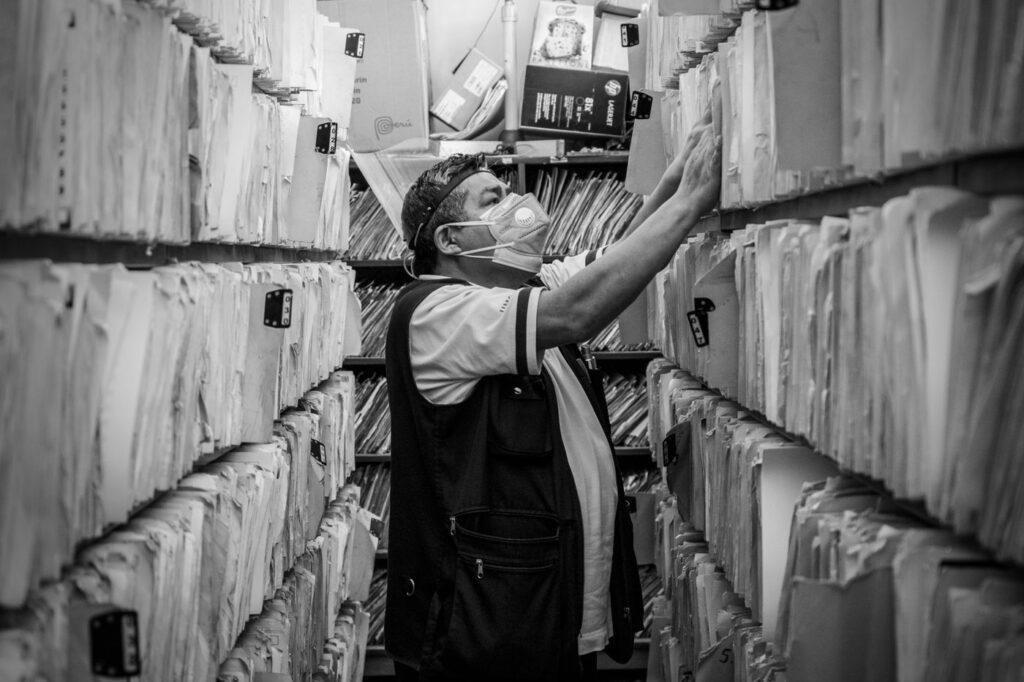
    The records are yours, and you do not have to explain why you want them
    The records are yours, and you do not have to explain why you want them.

    Request the radiographs specifically, in digital form if possible. Practices deal with these requests routinely, a reasonable copying fee may apply, and you do not owe anyone an explanation for wanting them. Federal guidance on getting a copy of your own health record sets out the general right of access that applies here.

    What to bring

    A useful second-opinion appointment vs. a wasted one
    ElementUsefulWasted
    RecordsRecent radiographs brought or sent aheadStarting again from scratch
    HistoryWhen it started, what triggers it, what has been tried“It has hurt for a while”
    FramingWhat are my options, and what happens if I wait“Was the last dentist wrong?”
    ExpectationA reasoned recommendation, possibly the same oneA guaranteed different answer
    TimingBefore anything irreversibleAfter the tooth is already out

    Swipe the table sideways to see every column.
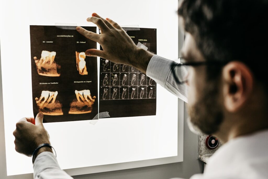
    Bringing recent images avoids repeat exposure and a repeated fee
    Bringing recent images avoids repeat exposure and a repeated fee.

    The framing row matters more than it looks. An appointment framed as a fault-finding exercise produces a defensive conversation; one framed around options produces a useful one.

    Who to ask

    Where you take the question matters as much as asking it. The right choice depends on what the disputed decision actually is.

    Where to take a second opinion
    The question is aboutBest placed to answer
    Whether a tooth can be root treated at allAn endodontist — this is the whole caseload
    Whether a failed root canal can be redoneAn endodontist, usually with 3D imaging
    Gum support and whether the tooth is stableA periodontist
    Implant planning and bone volumeAn oral surgeon or implant dentist
    An overall treatment plan across many teethA second general dentist

    Swipe the table sideways to see every column.

    The distinction to hold on to is that whoever provides the alternative treatment is not a neutral party on whether the alternative is needed. That is not a suggestion of bad faith — it is simply that everyone sees a case through what they do most.

    Questions worth asking

    Four questions get you most of the way, whether this is a first opinion or a second.

    What are my options, what happens if I wait, and what would you do?
    What are my options, what happens if I wait, and what would you do?
    1. What are my options, including doing nothing for now? There is nearly always more than one path.
    2. What happens if I wait three months? This separates urgent from elective, and the answer should be specific.
    3. How likely is this to work, and what does failure look like? A real prognosis comes with a number and a fallback.
    4. What would you do if this were your tooth? The most useful question in dentistry, and most clinicians will answer it honestly.

    If extraction has been proposed, add a fifth: what would it take to save this tooth, and what is the cost of trying? Comparing the alternatives is easier once both figures are on the table.

    Timing, and the one situation where it does not apply

    A second opinion is worth having before a decision, not after one. Once a tooth is out there is nothing left to assess, and once a crown has been cut the choice has largely been made.

    The exception is genuine urgency. If there is facial swelling, fever or spreading infection, that needs treating now — and the treatment for an acute abscess is not the part people disagree about. Get the infection under control, then take your time over the longer-term plan, which will still be there next week.

    Between those two extremes, most dental decisions tolerate a fortnight. If you are told a decision cannot wait a week and there is no swelling, no fever and no severe pain, that itself is worth a question.

    When the opinions agree

    This is the usual result, and it is not a wasted appointment. Two independent assessments reaching the same conclusion is exactly the reassurance you went looking for.

    Agreement is the common outcome, and it is worth having
    Agreement is the common outcome, and it is worth having.

    It also tends to change how the treatment feels. Going ahead with a plan you have examined and understood is a different experience from going ahead with one you were handed — which matters more than people expect if anxiety is part of the picture.

    When they disagree

    Disagreement in dentistry is usually about probability, not about facts. Both clinicians see the same crack; they differ on whether the tooth is worth attempting.
    The aim is a decision you would still make a year from now
    The aim is a decision you would still make a year from now.

    So ask each what they saw and what drove the recommendation, rather than treating it as a vote. Where the difference is about whether a difficult tooth can be treated at all, the opinion that carries more weight is generally the one with the equipment and the caseload to attempt it — which is why referral exists.

    Frequently asked questions

    Will my dentist be offended?

    Almost never. Second opinions are routine in every branch of healthcare, and most dentists would rather you were confident than quietly unconvinced. You do not need to explain your reasons.

    Do I have a right to my records?

    You have a right to a copy. The physical record belongs to the practice that created it, but professional guidance is clear that patients are entitled to copies, including radiographs. A reasonable copying fee may apply.

    Will I need new X-rays?

    Not if yours are recent. Bringing digital copies avoids a second dose and a second fee, which is the main practical reason to request them in advance.

    What if the two opinions disagree?

    Ask each what they saw that led them there. Disagreement is usually about the odds of saving a tooth rather than about the diagnosis, and hearing both reasonings is more useful than counting votes.

    Have Questions or Ready to Schedule?

    Our team is here to help — call, email, or request an appointment online.
    About this guide. Written for patients of Southeast Endodontics, PC, Charleston, SC. We see second-opinion consultations regularly, including cases where the answer is that the original plan was right.

  • Dental Emergency While Travelling: What to Do

    Dental Emergency While Travelling: What to Do

    Toothache eight hours from home is a logistics problem before it is a clinical one. What you need is a short list of what can wait, what cannot, and who to call.

    Here is how to sort the two, what belongs in your luggage, and how to find competent care in a place you do not know.

    Key takeaways

    • Swelling, fever or difficulty swallowing needs care that day, wherever you are.
    • A lost crown or filling can usually be managed for a few days with pharmacy supplies.
    • Most domestic dental plans stop at the border; travel policies typically cover pain relief only.
    • A check-up before a long trip is the cheapest insurance available.

    Sort it first: wait, or find someone today

    The distinction that matters is not how much it hurts. It is whether infection is spreading.

    Manage the pain, but do not assume it will hold until you fly home
    Manage the pain, but do not assume it will hold until you fly home.
    What can wait and what cannot
    SituationWhat to do
    Facial swelling, fever, difficulty swallowing or openingEmergency care that day — do not wait for the flight home
    Severe pain keeping you awakeFind a dentist locally; this rarely improves on its own
    Tooth knocked outWithin the hour — keep it moist, do not scrub it
    Crown or filling lost, no painManageable for days with pharmacy temporary cement
    Mild sensitivity to hot or coldUsually holds; sensitive toothpaste and avoid the trigger
    Chipped tooth, no sharp edge, no painWait until you are home

    Swipe the table sideways to see every column.

    The top row is the one people misjudge. Swelling that spreads toward the eye or down the neck is a medical emergency, not a dental inconvenience — the abscess warning signs are the same abroad as at home.

    What to pack

    A pouch that fits in a wash bag covers the majority of travel dental problems, and everything in it is available before you leave.

    A small kit handles most of what goes wrong
    A small kit handles most of what goes wrong.
    • Temporary filling material or dental cement from a pharmacy — for a lost filling or a crown that has come off.
    • Ibuprofen and paracetamol, which work better alternated than either alone.
    • Orthodontic wax, which covers a sharp edge and stops it shredding your tongue.
    • A small container with a lid — useful if a crown or a tooth comes out.
    • Your dentist’s contact details saved offline, not only in an email you need signal to open.

    If you already have a temporary restoration in place, take the material rather than hoping. Temporaries are designed to last weeks, and a long trip is exactly when one gives way.

    Before you go

    If you have a symptom you have been ignoring, deal with it before a long trip rather than after. A tooth that twinges at home reliably becomes the tooth that keeps you awake in a hotel room.

    The cheapest travel insurance is a check-up before you leave
    The cheapest travel insurance is a check-up before you leave.

    This applies particularly to anything already under way — a temporary crown, an unfinished root canal, a tooth that has been sensitive for weeks. Pain on biting is the classic symptom people postpone, and it does not improve with distance.

    Finding a dentist you do not know

    Do not simply walk into the nearest practice. Four sources are better than a map search, in roughly this order.

    Hotel desks, consulates and insurers all keep lists of English-speaking dentists
    Hotel desks, consulates and insurers all keep lists of English-speaking dentists.
    Where to look, and what each is good for
    SourceUseful for
    Your travel insurerA vetted clinic, and confirmation of what is covered before you commit
    Your embassy or consulateLists of English-speaking practitioners; the standard first port of call
    Hotel concierge or receptionFast local knowledge; they deal with this often
    A local hospital emergency departmentSwelling, fever, or anything systemic

    Swipe the table sideways to see every column.

    Ask two questions when you arrive: what exactly are you doing today, and what will it cost. Emergency treatment abroad should stabilise the problem — relieve pain, drain infection, place a temporary. It is rarely the moment for a full course of treatment you cannot follow up.

    Ask your own dentist from where you are

    This is underused. A clear photograph and a description of the symptoms is often enough for someone who knows your mouth to tell you whether this waits.

    A photograph and a phone call can settle whether it can wait
    A photograph and a phone call can settle whether it can wait.

    They also know what is already going on in that tooth, which a stranger does not. If you have had a root canal recently and the tooth is suddenly painful again, that history changes the assessment considerably — see what a returning symptom usually means.

    The insurance trap

    Two gaps catch people out. Domestic dental insurance generally does not travel. And travel insurance usually covers emergency pain relief only, with definitions that exclude most actual treatment.

    Most domestic dental plans stop at the border
    Most domestic dental plans stop at the border.

    Read the dental section before you leave, keep every receipt and any radiographs, and expect to pay at the time and claim afterwards. Cost and coverage covers how this works at home for comparison.

    Managing pain until you are seen

    Whatever the plan, you may have hours to get through first. A few things help and one common mistake makes matters worse.

    Alternating ibuprofen and paracetamol works better than either at double dose, and staying ahead of the pain is more effective than chasing it. Cold against the cheek helps swelling; heat generally does not, and can encourage an infection to spread.

    Do not place aspirin against the gum. It burns the tissue without touching the tooth, and it remains one of the commonest self-treatments we see the aftermath of. If you are already carrying antibiotics from home, do not start them on your own judgement — they treat spreading infection, not toothache, and taking a partial course makes the eventual treatment harder.

    When you get back

    Have anything done abroad checked. A temporary placed in another country is still a temporary, and it needs replacing on the same timescale as one placed down the road.

    Bring whatever paperwork and images you were given. The American Association of Endodontists’ guide to dental symptoms and what they signal is a reasonable reference for judging severity while you are away.

    Frequently asked questions

    Does my dental insurance cover me abroad?

    Usually not. Most domestic dental plans stop at the border, and most travel insurance covers emergency pain relief only — not crowns, root canals or anything described as routine. Check the wording before you go rather than at the counter.

    Can a root canal wait until I get home?

    Sometimes, if you are days rather than weeks from returning and there is no swelling. Swelling, fever or spreading pain never waits. If in doubt, send your own dentist a photograph and describe the symptoms.

    Is dental care abroad safe?

    Standards vary by country and by practice rather than by continent. Ask about sterilisation, use single-use items where offered, and prefer a clinic recommended by your insurer, consulate or hotel over one found by walking past it.

    What if a crown comes off while I am away?

    Keep it. Do not glue it back with household adhesive. A pharmacy temporary cement will usually hold it for a few days, and your own dentist can re-cement it properly on your return.

    Have Questions or Ready to Schedule?

    Our team is here to help — call, email, or request an appointment online.
    About this guide. Written for patients of Southeast Endodontics, PC, Charleston, SC. If you are a patient of ours and something goes wrong while travelling, call the practice — we would rather advise you from a distance than see the result later.

  • Why Isn’t My Tooth Healing? What Affects the Outcome

    Why Isn’t My Tooth Healing? What Affects the Outcome

    A root canal is not a repair you can inspect. Most of what determines whether it worked happens over the following months, in bone you cannot see, influenced by things that have nothing to do with the appointment itself.

    Here is what actually governs endodontic healing, which factors are within your control, and how success is genuinely judged.

    Key takeaways

    • Bone at the root tip rebuilds over six to twelve months — symptoms resolve far sooner.
    • Diabetes and smoking are both established negative prognostic factors.
    • The quality of the restoration above the root filling predicts survival as strongly as the root filling itself.
    • Healing is judged by comparing radiographs over time, not by how the tooth feels.

    Two timelines, not one

    Confusion here is almost universal, and it comes from conflating two separate processes.

    Bone at the root tip rebuilds over six to twelve months, not weeks
    Bone at the root tip rebuilds over six to twelve months, not weeks.

    Symptoms resolve quickly. Once infected tissue is removed and pressure released, most people are comfortable within days — the recovery guide covers that week in detail.

    Bone healing is slow. Where infection destroyed bone around the root tip, that defect fills in gradually over six to twelve months, sometimes longer for a large lesion. A tooth that feels perfect at three months may still show a shadow, and that is expected rather than alarming.

    What is outside your control

    Prognostic factors established before treatment starts
    FactorEffectWhy
    Size of the pre-existing lesionLarger lesions heal more slowlyMore bone to rebuild
    Whether this is a retreatmentLower success than first-time treatmentEstablished biofilm is harder to remove
    Root anatomyCurved or calcified canals are harder to clean fullyAccess limits what any technique can reach
    A crack in the toothOften decisiveA route bacteria can keep using
    Existing bone supportPoor support limits the ceilingThe tooth may be sound and still not viable

    Swipe the table sideways to see every column.

    None of those are anyone’s fault, and they are why an honest prognosis comes with a percentage attached rather than a promise. A tooth with a large lesion and a previous failed treatment is a different proposition from a straightforward first-time case.

    What is within your control

    Systemic health genuinely affects periapical healing, and this is where the evidence has firmed up over the last decade.

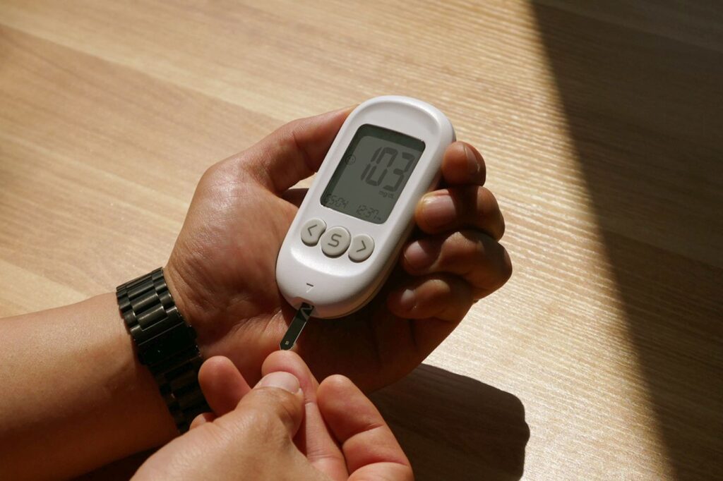
    Diabetes is an established prognostic factor in endodontic healing
    Diabetes is an established prognostic factor in endodontic healing.

    Diabetes is the best-documented factor. Systematic reviews associate it with poorer healing, more persistent lesions and a higher proportion of teeth not retained. Control matters more than diagnosis — well-managed diabetes behaves very differently from poorly managed diabetes, and it is worth mentioning at the consultation rather than leaving it off the medical history.

    Where diabetes is part of the picture, it changes what we tell you rather than whether we treat. Expect a longer review interval before anyone calls the tooth healed, and expect more attention paid to the gums around it, because periodontal and periapical problems tend to travel together.

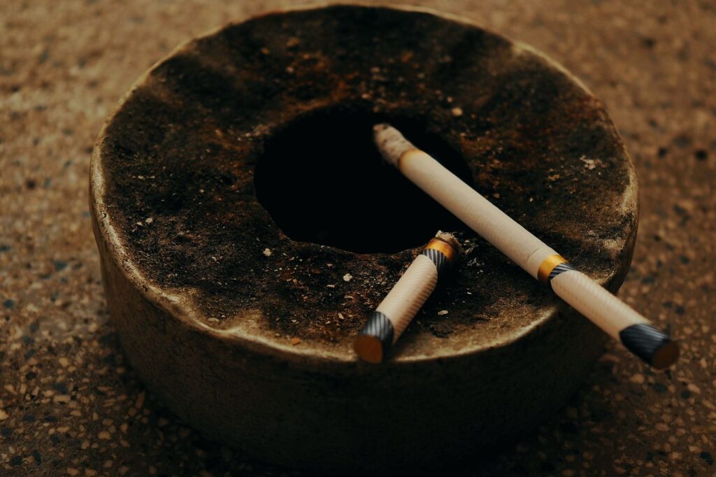
    Smoking is associated with poorer healing and more retained lesions
    Smoking is associated with poorer healing and more retained lesions.

    Smoking is the second. It reduces blood supply to the tissues doing the healing, and studies of apical periodontitis after treatment show measurably worse resolution in smokers. Because healing continues for months, cutting down after treatment still helps.

    The factor people never think about

    Ask what causes root canals to fail and most people say the root canal was done badly. The evidence points somewhere else: the commonest route is recontamination from above.

    The quality of the restoration above predicts survival as much as the root filling
    The restoration above predicts survival as strongly as the root filling.

    A leaking filling, a temporary left too long, or a crown that never got placed lets bacteria back into canals that were properly cleaned. Studies of long-term survival consistently find restoration quality to be as strong a predictor as obturation quality.

    That is genuinely within your control — it means booking the permanent restoration promptly. On a back tooth that means a crown, and the temporary is not a destination.

    What normal looks like at each stage

    Because the two timelines run at different speeds, it helps to know roughly what should be true when.

    The normal course after root canal treatment
    Point in timeWhat is usualWhat is not
    First 2–3 daysTenderness to bite, easing dailySwelling, fever, or pain that worsens
    1–2 weeksComfortable; permanent restoration bookedPain unchanged from day one
    1–3 monthsNo symptoms; tooth functioning normallyA gum boil, or discomfort returning
    6 monthsRadiograph shows a shrinking lesionA lesion the same size or larger
    12 monthsLesion largely or fully resolvedNo change — time to reassess options

    Swipe the table sideways to see every column.

    The middle rows are where people either relax too early or worry too much. A tooth that is quiet at three months is behaving normally even though the radiograph will still show something; a tooth that is uncomfortable at three months is worth reporting rather than waiting out.

    How healing is actually judged

    Not by symptoms. A radiograph taken at six or twelve months is compared with the one taken before treatment, and what matters is the direction: a shrinking lesion is healing, whatever the tooth feels like.

    Comparison over time is how healing is actually judged
    Comparison over time is how healing is judged.

    This is why review appointments exist and why they are easy to skip. A tooth that is comfortable but shows an unchanged lesion at twelve months is precisely the case worth catching — the options at that point, covered in retreatment, surgery or extraction, are far better than the options two years later.

    What to do with all this

    Three things make a measurable difference: get the permanent restoration placed promptly, manage the systemic factors you can, and attend the reviews even when nothing hurts.

    Some factors are fixed; several are genuinely within your control
    Some factors are fixed; several are genuinely within your control.

    None of that is dramatic, which is rather the point. Endodontic outcomes are decided by unglamorous things — a crown fitted on time, blood sugar in range, a radiograph taken at six months — far more often than by anything that happens in the chair.

    And give it time. The single commonest reason people believe a root canal has failed is judging it at eight weeks against a process that takes eight months. The British Dental Journal’s review of success and failure in endodontic treatment sets out the predictors in clinical detail.

    Frequently asked questions

    How long should healing take?

    Symptoms usually settle within days to a couple of weeks. Bone at the root tip is far slower — six to twelve months is normal, and larger lesions can take longer. Judging success at three months is judging it too early.

    Does diabetes mean my root canal will fail?

    No, but it is an established prognostic factor. Well-controlled diabetes has much less effect than poorly controlled diabetes, and the practical consequence is usually a longer follow-up before we call it healed.

    Will quitting smoking help now, after treatment?

    It helps. Healing continues for months after the appointment, so reducing or stopping during that window still affects the outcome rather than only mattering beforehand.

    My tooth feels fine. Does it still need reviewing?

    Yes. Comfort and healing are different things — a comfortable tooth can still show an unresolved lesion on a radiograph, and that is exactly the case worth catching early.

    Have Questions or Ready to Schedule?

    Our team is here to help — call, email, or request an appointment online.

    About this guide. Written for patients of Southeast Endodontics, PC, Charleston, SC. Prognostic factors describe populations rather than individuals — your own tooth is assessed on its own findings.

  • Root Canals in Older Adults: What Changes and What Doesn’t

    Root Canals in Older Adults: What Changes and What Doesn’t

    People in their seventies and eighties routinely ask whether a root canal is worth it at their age. The evidence answers that clearly: age does not affect whether treatment works.

    What does change is how the tooth behaves and what else is going on medically. Here is what genuinely differs about endodontic treatment later in life.

    Key takeaways

    • Across two dozen studies, age is not a prognostic factor for root canal outcome.
    • Canals narrow throughout life, which makes treatment technically harder rather than less successful.
    • Medications matter far more than age in planning treatment.
    • Dry mouth from medication is the commonest driver of new decay later in life.

    What actually changes with age

    Teeth lay down dentine continuously. Every year the pulp chamber gets a little smaller and the canals a little narrower, and by seventy a canal that was straightforward at twenty can be extremely fine.

    Canals narrow over a lifetime as secondary dentine is laid down
    Canals narrow over a lifetime as secondary dentine is laid down.

    The pulp itself changes too: more fibrous tissue, less blood supply, fewer nerve fibres. That has a practical consequence — an older tooth often responds more weakly to cold testing, and a reduced response is not necessarily a dead pulp. It has to be read against the neighbouring teeth rather than in isolation.

    Why calcified canals are harder, not hopeless

    A calcified canal is not blocked. It is narrow, and the entrance is often hidden under a layer of dentine laid down over decades.

    Finding a calcified canal is a magnification problem more than anything
    Finding a calcified canal is largely a magnification problem.

    Locating it is a magnification and imaging problem more than a mechanical one. Under a microscope the subtle colour change marking a canal entrance is visible; without one it frequently is not. 3D imaging shows whether a canal exists and where it runs before any drilling starts, which matters when the alternative is exploring blind.

    Appointments take longer as a result. That is worth knowing in advance rather than being surprised by it, and it is one of the clearer reasons a general dentist may prefer to refer a heavily calcified tooth rather than attempt it — see what usually prompts a referral. The tooth is not more likely to fail; it simply needs equipment and time that a routine appointment does not allow for.

    What matters more than age

    Factors that genuinely affect planning
    FactorWhy it mattersWhat we do
    AnticoagulantsBleeding during surgical proceduresUsually continued; coordinated with your physician
    Bisphosphonates or denosumabAffect bone healing after extractionOften makes saving the tooth preferable to removing it
    Dry mouth from medicationSharply raises decay riskPrevention advice, and expect more frequent review
    DiabetesSlower periapical healingLonger follow-up before judging success
    Difficulty lying backComfort and breathingShorter appointments, adjusted chair position
    Limited mouth openingAccess to back teethPlanned in advance rather than discovered

    Swipe the table sideways to see every column.

    Medication lists matter more than age in planning treatment
    Medication lists matter more than age in planning treatment.

    The bisphosphonate row is the one people underestimate. If you take or have taken these drugs, extraction carries a small but real risk to the jawbone — which shifts the balance firmly toward keeping the tooth. That reverses the usual assumption that pulling is the simpler option.

    Comfort during a longer appointment

    If the technical work takes longer, the practical question becomes whether you can comfortably stay in the chair for it. That is worth raising at the consultation rather than enduring on the day.

    Difficulty lying flat is the commonest issue, whether from breathing, reflux or a bad back. The chair position can be adjusted, and treatment can be divided across two shorter visits instead of one long one. Neither compromises the result.

    Anaesthetic works no differently with age, though it can take slightly longer to wear off. If you take medication for blood pressure or heart rhythm, tell us — it occasionally changes which anaesthetic we choose, and it is easier to plan for than to discover. Sedation options remain available and are assessed against your medical history rather than ruled out by age.

    Decades of previous dentistry

    Older teeth rarely arrive untouched. Crowns, posts, large fillings and previous root canals all change the approach.

    Decades of previous dentistry change how a tooth is accessed
    Decades of previous dentistry change how a tooth is accessed.

    Reaching the pulp through an existing crown means cutting through it, and the crown may or may not survive. Where a post is present, removing it carries a risk of fracturing the root — which is one of the situations where root-end surgery is preferred over conventional retreatment.

    Dry mouth, and why it causes so much trouble

    Hundreds of common medications reduce saliva — blood pressure drugs, antidepressants, antihistamines, diuretics. Saliva buffers acid, washes away debris and carries minerals that repair early damage, so losing it changes the mouth substantially.

    Dry mouth from medication is the commonest driver of decay later in life
    Dry mouth from medication is the commonest driver of decay later in life.

    The characteristic result is decay at the gumline and around the edges of old restorations, in people who went decades without a cavity. It is worth raising with whoever prescribes the medication and worth managing actively, because it is the commonest route to needing a root canal at seventy.

    Is it worth treating?

    This is the question behind the question, and it deserves a direct answer. Success rates are the same; a treated tooth lasts as long as any other; and the alternatives have their own costs.

    Keeping natural teeth into later life is now the expectation, not the exception
    Keeping natural teeth into later life is now the expectation, not the exception.

    Extraction means either a gap that affects chewing, or a replacement. Implants take months and need adequate bone; dentures change what you can eat. The alternatives compared covers the trade-offs, and the calculation frequently favours keeping the tooth more strongly at seventy than at forty.

    Saving the tooth vs. replacing it, later in life
    ConsiderationRoot canal and crownExtraction and implant
    Time to a finished resultWeeksSeveral months
    Number of appointmentsTypically two to threeConsiderably more
    Depends on bone volumeNoYes — may need grafting first
    Affected by bisphosphonate historyNoYes, and significantly
    Surgical procedure requiredUsually noneYes
    Keeps the natural root and ligamentYesNo

    Swipe the table sideways to see every column.

    The time column is the one that decides it for many people. Months of a gap, a temporary and repeated visits is a real cost when the alternative finishes in a fortnight.

    Published work on endodontic considerations in older patients covers the clinical detail behind this.

    Frequently asked questions

    Am I too old for a root canal?

    No. Across two dozen studies, patient age does not affect the outcome of root canal treatment. The technical demands can be higher, but the results are not worse.

    Why does my dentist say the canals are calcified?

    Because they narrow throughout life. Teeth lay down dentine continuously, so a canal that was wide at twenty can be very fine at seventy. It makes the tooth harder to treat, not impossible.

    I take several medications. Does that matter?

    It matters more than your age does. Anticoagulants, bisphosphonates and drugs causing dry mouth all affect planning, so bring a current list to the appointment.

    Is extraction simpler at my age?

    Not necessarily. Older bone is denser and roots can be more brittle, so extraction is not automatically the easier option — and replacing the tooth afterwards has its own considerations.

    Have Questions or Ready to Schedule?

    Our team is here to help — call, email, or request an appointment online.

    About this guide. Written for patients of Southeast Endodontics, PC, Charleston, SC. Bring a current medication list and your physician’s details to any appointment — they change planning more than anything else here.

  • Can the Nerve Be Saved? Vital Pulp Therapy in Adults

    Can the Nerve Be Saved? Vital Pulp Therapy in Adults

    For decades, a pulp inflamed past a certain point meant one thing: remove all of it. That position has genuinely shifted, and it is one of the more interesting changes in endodontics.

    Vital pulp therapy keeps living pulp tissue in the tooth rather than removing it entirely. Here is what it involves, the evidence behind it, and the cases where a conventional root canal remains the right answer.

    Key takeaways

    • Only the inflamed part of the pulp is removed; healthy tissue in the roots stays alive.
    • Trials in mature teeth report success comparable with root canal treatment — around 93% in several studies.
    • Less post-operative pain on day one, shorter appointments and lower cost.
    • Candidacy is decided during the appointment, based on how the pulp actually behaves.

    What it actually is

    A conventional root canal removes the pulp from the crown and every root, then fills the space. Vital pulp therapy removes only the part that is inflamed — usually the portion in the crown — and seals a medicated material over the healthy tissue beneath.

    A pulpotomy removes the inflamed crown portion and leaves the roots
    Only the inflamed portion is removed; the rest of the pulp stays alive.

    The reasoning is that pulp inflammation is often localised rather than uniform. The tissue nearest the decay may be beyond saving while the tissue deeper in the roots is perfectly healthy, and removing all of it treats the whole pulp for a problem confined to part of it.

    Why this is newer than it sounds

    Partial pulp treatment is not a new idea. What changed is the materials.

    Modern bioceramic materials are what made this predictable
    Modern bioceramic materials are what made the approach predictable.

    Older capping materials sealed poorly and irritated the tissue they were meant to protect, so results were unreliable and the approach fell out of favour in adults. Hydraulic calcium silicate cements seal well, tolerate moisture, and actively stimulate the pulp to lay down a hard tissue barrier of its own. That is the difference between an optimistic technique and a predictable one.

    What the evidence shows

    Vital pulp therapy vs. root canal treatment in mature teeth
    MeasureFull pulpotomyRoot canal treatment
    Clinical success~97%~99%
    Radiographic success~93%~95%
    Pain on day one after treatmentSignificantly lowerHigher
    Treatment timeShorterLonger
    CostLowerHigher
    Patient satisfactionHigher across time, pain and costLower on those measures

    Swipe the table sideways to see every column.

    Those figures come from randomised trials comparing the two in mature teeth with symptomatic irreversible pulpitis — the exact diagnosis that used to mean an automatic root canal. Longer-term series using calcium silicate materials report success between roughly 78% and 90% at one to five years.

    The honest caveat is that the follow-up periods are shorter than for conventional treatment, which has decades of data behind it. This is well-supported rather than fully settled.

    Who it suits

    The typical case is a tooth where decay has reached the pulp and there are symptoms, but the tooth is otherwise sound and restorable.

    It applies where decay has reached the pulp but the tissue can still recover
    It applies where decay has reached the pulp but the tissue can still recover.

    Candidacy is genuinely decided in the chair. Once the decay is removed and the pulp exposed, what matters is how it bleeds: healthy pulp bleeds bright red and stops within a few minutes under gentle pressure. Bleeding that will not stop indicates inflammation deeper than the part being removed, and treatment converts to a conventional root canal in the same appointment.

    When each approach applies
    SituationUsual approach
    Decay reaching the pulp, bleeding controls quicklyVital pulp therapy
    Bleeding will not stop after several minutesConvert to root canal treatment
    Pulp already dead — no response to coldRoot canal treatment
    Swelling or an abscess presentRoot canal treatment
    Tooth needs a crown for structural reasons anywayOften root canal treatment
    Immature tooth in a child or teenagerVital pulp therapy strongly preferred

    Swipe the table sideways to see every column.

    That last row is long-established practice rather than a new development — see endodontics in children, where keeping the pulp alive lets the root finish forming.

    Why keeping the pulp is worth something

    A tooth with a living pulp retains sensation, which means it can still tell you when something is wrong. A root-filled tooth cannot, so problems there are found on radiographs rather than reported as symptoms.

    A living pulp keeps sensing and defending the tooth
    A living pulp keeps sensing and defending the tooth.

    Living pulp also continues laying down dentine in response to wear and irritation, and less tooth structure is removed in the first place — which matters for long-term fracture resistance, as the survival data on cuspal coverage shows.

    What the appointment is like

    It starts the same way as any other treatment: local anaesthetic, a rubber dam, and the decay removed completely. The difference comes at the point the pulp is reached.

    Instead of proceeding into the roots, the exposed tissue is removed to a defined level and bleeding is controlled under light pressure. That pause is the diagnostic step — what happens in those few minutes decides the rest of the appointment.

    If bleeding settles, the material is placed directly against the remaining pulp and sealed, and the tooth is restored in the same visit. Post-operative discomfort is typically milder than after conventional treatment.

    What it does not change

    The restoration afterwards is as important as it is following a root canal. A well-treated pulp under a leaking filling will fail, because bacteria simply return.

    Shorter appointment, lower cost, and less post-operative pain on day one
    Shorter appointment and lower cost, but the restoration still matters.

    Follow-up matters too. These teeth are reviewed for symptoms and for continued pulp response, and a proportion do go on to need conventional treatment later — which is a manageable outcome rather than a disaster.

    Asking about it

    It is a reasonable question to raise if you have been told you need a root canal on a tooth that is otherwise in good condition. The answer may well be no — but the reasoning is worth hearing.

    Where the pulp is too far gone, conventional treatment remains the answer
    Where the pulp is too far gone, conventional treatment remains the answer.

    What it is not is a way to avoid treatment. A tooth with a dead pulp or an abscess needs conventional root canal treatment, and delaying in the hope of a lesser option makes that worse. The published randomised trial comparing full pulpotomy with root canal therapy is the source for the figures above.

    Frequently asked questions

    Is this the same as the pulp capping done on children?

    The principle is the same — keep living pulp alive — but the evidence in adults is newer. What changed is the materials: hydraulic calcium silicate cements seal and stimulate repair far better than the older options, which is why adult cases now succeed at rates comparable with root canal treatment.

    How would I know if I am a candidate?

    It is decided at the appointment, not before. The deciding factor is what the pulp looks like once the decay is removed and how it bleeds. If bleeding stops within a few minutes under pressure, the tissue is usually healthy enough.

    What happens if it does not work?

    You have a conventional root canal, which was the alternative anyway. Failure means treatment moves on rather than the tooth being lost, and that is the reason it is reasonable to attempt.

    Is it cheaper?

    Usually, yes — published trials report lower cost and shorter treatment time than root canal therapy. It still needs a proper restoration afterwards, which is the larger part of the bill on a back tooth.

    Have Questions or Ready to Schedule?

    Our team is here to help — call, email, or request an appointment online.

    About this guide. Written for patients of Southeast Endodontics, PC, Charleston, SC. Vital pulp therapy is well supported but has shorter follow-up data than conventional treatment; whether it suits your tooth is decided clinically.

  • Temporary Fillings: Looking After a Tooth Mid-Treatment

    Temporary Fillings: Looking After a Tooth Mid-Treatment

    Between the appointment where a root canal is started and the one where the permanent restoration goes on, a small grey or white plug of material is the only thing protecting the work underneath. It deserves more attention than it usually gets.

    Here is what a temporary filling is actually doing, how to look after it, and what to do when it comes out.

    Key takeaways

    • The temporary seals cleaned canals against saliva — that seal is what makes the treatment last.
    • They are designed for weeks, not months.
    • A lost temporary is a same-week phone call, not an emergency, but do not leave it.
    • Chew on the other side and avoid hard or sticky food until the permanent restoration is fitted.

    What it is actually doing

    Reaching the canals means cutting an opening through the biting surface. That opening has to be closed between visits, and the closure has two jobs.

    It seals the access cavity and keeps saliva out of cleaned canals
    It seals the access cavity and keeps saliva out of cleaned canals.

    The first is sealing. Saliva carries bacteria, and canals that have been cleaned and disinfected are recontaminated within days if saliva reaches them. That single fact is why the temporary matters — leakage under a temporary is a recognised route to treatment failing later.

    The second is being removable. A permanent material would have to be drilled out, removing more tooth structure each time. Temporary materials seal well and come away cleanly, which is exactly the trade-off you want mid-treatment.

    Why treatment sometimes takes two visits

    Most root canals are completed in one appointment. Where they are not, it is usually deliberate rather than a shortfall of time — our guide to what happens during treatment covers the sequence.

    Between visits the canal usually holds an antimicrobial dressing
    Between visits the canal usually holds an antimicrobial dressing.

    Where there is significant infection, an antimicrobial dressing is placed in the canal and left to work for a week or two. Sealing an actively discharging canal traps the problem inside, so waiting is the safer choice. The temporary holds that dressing in place.

    Looking after it

    Living with a temporary filling
    DoDo not
    Chew on the opposite sideTest it by biting on that tooth
    Brush normally, gently over the toothSkip brushing the area because it feels fragile
    Floss, pulling the strand out sidewaysSnap floss up out of the contact — it lifts temporaries
    Eat soft food for the first dayEat nuts, ice, crusty bread, toffee or gum
    Expect mild tenderness for a few daysIgnore pain that is increasing after day three
    Keep the next appointmentLet it run for months because the tooth feels fine

    Swipe the table sideways to see every column.

    Flossing is the one people get wrong. Do floss — leaving the area unclean causes its own problems — but pull the strand out sideways rather than snapping it up through the contact, which can lift the temporary out with it.

    If it comes out

    1. Call the practice. Same day if possible, next working day otherwise. Replacing it takes five to ten minutes and usually needs no injection.
    2. Rinse gently with warm salt water — about half a teaspoon in a glass — to clear debris.
    3. Keep chewing away from it entirely until it is resealed.
    4. Brush around it carefully rather than avoiding the area.
    5. Do not poke at the opening with anything, and do not try to clean inside it.
    A lost temporary is not an emergency, but it is a same-week phone call
    A lost temporary is not an emergency, but it is a same-week phone call.

    If you genuinely cannot be seen for several days, an over-the-counter temporary repair material can bridge the gap. Use it as a short-term measure only, and tell us you have used it — it changes what we find when we reopen the tooth.

    Rinse gently with warm salt water and keep the area clean
    Rinse gently with warm salt water and keep the area clean.

    What is normal between visits, and what is not

    Mild tenderness, particularly when biting on that side, is expected and normal. The ligament around the root has been inflamed both by the original problem and by the treatment itself, and it settles over several days — our recovery guide covers the timeline in detail.

    What is not normal: pain increasing after the third day, facial swelling, fever, or a bad taste appearing from the tooth. Those warrant a call rather than waiting for the scheduled appointment — see which symptoms need same-day care.

    A tooth that feels completely normal is also not a reason to skip the next visit. Comfort returns long before the tooth is finished, and that gap is precisely when people drift.

    How long you actually have

    Temporary materials are not all the same, and the timescale depends on what was placed and where.

    Temporary restorations and how long each is designed to last
    SituationDesigned to lastRisk if left longer
    Soft temporary between two treatment visits1–3 weeksWears through; dressing leaks out
    Firmer temporary after treatment is completedSeveral weeksMarginal leakage into the canals
    Temporary crown on a prepared toothWeeksDebonds; the tooth can drift
    Any temporary on a back toothWeeksCusp fracture under chewing load
    Any temporary, months onNot designed for itRetreatment, or loss of the tooth

    Swipe the table sideways to see every column.

    The bottom row is not scaremongering. Teeth that are treated and then never permanently restored are a recognisable category in every endodontic practice, and they usually arrive back with either a fractured cusp or a reinfected canal.

    The deadline nobody mentions

    This is the part worth taking away. A temporary filling is a component with a service life measured in weeks.

    The temporary is a bridge to the permanent restoration, not a destination
    The temporary is a bridge to the permanent restoration, not a destination.

    Left for months it wears down under chewing and begins to leak, and bacteria re-enter canals that were cleaned at some expense. The tooth then needs retreating — not because the root canal was poor, but because the seal above it failed.

    On a back tooth the permanent restoration also means a crown rather than a filling, and outcomes are measurably better when it follows within about sixty days. Book that appointment before you leave, not once something starts bothering you again. A long trip is exactly when a temporary gives way, which is why it features in planning for dental problems abroad. The ADA’s overview of root canal treatment covers the wider sequence.

    Frequently asked questions

    My temporary filling came out. Is it an emergency?

    Not an emergency, but not something to leave either. Call the same day or the next working day. The canal underneath has been cleaned and needs to stay sealed, and an open access cavity lets saliva and bacteria straight back in.

    Can I use a drugstore temporary filling kit?

    As a short bridge if you genuinely cannot be seen quickly, yes, following the packet. If you can be seen within a day or two it is better to leave it alone — material packed into an access cavity can be awkward to remove cleanly.

    How long is a temporary filling meant to last?

    Weeks, not months. They are designed to seal reliably between appointments and then be removed easily. Left for months they wear, leak, and undo the work underneath.

    Can I eat normally with one?

    Chew on the other side. Hard and sticky foods are the two that dislodge temporaries — nuts, ice, crusty bread, toffee and chewing gum are the usual culprits.

    Have Questions or Ready to Schedule?

    Our team is here to help — call, email, or request an appointment online.
    About this guide. Written for patients of Southeast Endodontics, PC, Charleston, SC. If a temporary has come out and you are unsure how urgent it is, call and ask — that question takes a minute to answer.

  • What a 3D Scan Shows That an X-Ray Cannot

    What a 3D Scan Shows That an X-Ray Cannot

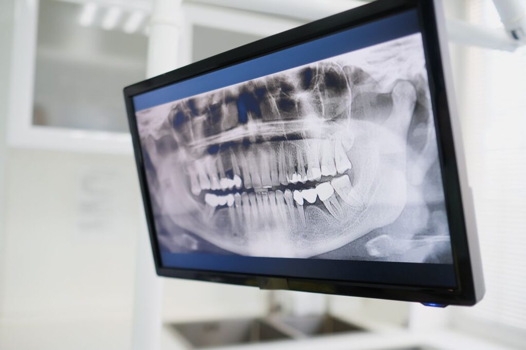
    A standard dental X-ray compresses a three-dimensional root into a flat picture. Most of the time that is enough. Occasionally the thing you most need to see is hidden precisely because of that flattening.

    Here is what cone-beam imaging shows that a film cannot, when it is genuinely worth taking, and why it should not be routine.

    Key takeaways

    • A conventional radiograph superimposes structures; a scan separates them into three planes.
    • A limited field-of-view scan costs roughly the dose of two periapical radiographs.
    • It is used selectively — the professional position is explicitly against routine use.
    • The commonest findings are extra canals, resorption, and the true extent of infection.

    What a flat image hides

    A periapical radiograph is a shadow. Everything between the source and the sensor is projected onto one plane, so structures sitting behind one another overlap.

    A standard radiograph compresses a three-dimensional root into a flat image
    A standard radiograph compresses a three-dimensional root into a single plane.

    In a molar with four canals, two frequently lie directly behind the others from the angle the image is taken. They are not faint on the film — they are invisible, hidden behind the roots in front. Bone loss also has to reach a certain extent before it registers at all, so early changes at a root tip can be genuinely undetectable.

    What a scan adds

    Cone-beam imaging takes a series of images as the machine rotates, then reconstructs them into a volume that can be viewed in cross-section from any angle.

    An extra canal hidden behind another is the classic 3D finding
    An extra canal hidden behind another is the classic 3D finding.
    What each type of imaging shows
    QuestionStandard radiograph3D scan
    How many canals does this tooth have?Often incompleteReliable, including canals hidden behind others
    How far has the infection spread?Underestimates early lesionsShows true extent in three dimensions
    Is there resorption, and how far around?Frequently missedShows position and circumferential spread
    Where is the sinus or nerve relative to the root?ApproximatePrecise — important before surgery
    Is this root fractured?Rarely visibleSometimes visible; still not guaranteed
    Routine diagnosis of a straightforward toothEntirely adequateUnnecessary

    Swipe the table sideways to see every column.

    That last row matters as much as the others. For most teeth a conventional radiograph answers the question completely, and taking a scan adds dose without adding information.

    When it is actually indicated

    The joint position of the endodontic and oral radiology specialties is that scans should be used selectively, guided by the clinical question. In practice that means:

    A limited field scan covers a few teeth at a far smaller dose
    A limited field scan covers a few teeth at a far smaller dose than a full-head study.
    • Retreatment, where the reason the first attempt failed is not visible — see why root canals fail.
    • Suspected extra canals, particularly in upper molars.
    • Suspected resorption, where extent decides whether the tooth is restorable — see how resorption is assessed.
    • Before surgery, to establish exactly where the sinus floor or the nerve to the lip sits relative to the root tip.
    • Dental trauma, where root fractures and displacement are hard to assess on a film.
    • Persistent symptoms with no explanation on conventional imaging.

    What we actually find

    Scans are worth taking only where the finding changes something. In endodontic practice a handful of findings recur.

    Common scan findings and what each changes
    FindingHow often it mattersWhat changes
    An untreated canal in a previously treated toothThe commonest useful findingRetreatment becomes worthwhile instead of extraction
    A lesion larger than the film suggestedFrequentPrognosis and follow-up interval
    Resorption, and how far around the rootOccasional but decisiveWhether the tooth is restorable at all
    Root tip within or against the sinusCommon in upper molarsSurgical approach, and what we warn you about
    Nerve canal close to a lower rootCommon in lower molarsWhether surgery is advisable
    Nothing unexpectedOftenConfirms the original plan — still useful

    Swipe the table sideways to see every column.

    The last row deserves as much weight as the first. A scan that confirms there is nothing hidden is not a wasted scan; it converts a suspicion into a decision, and it is frequently what allows treatment to proceed with confidence rather than caution.

    It also occasionally shows something outside the tooth entirely — sinus disease, or a lesion unrelated to the tooth we were investigating. Where that happens we refer it on rather than treating around it.

    The dose question, answered properly

    A scan is more radiation than a single film. That is worth stating plainly rather than glossing.

    Scans are taken selectively, never routinely — the ALARA principle
    Scans are taken selectively, never routinely — the ALARA principle.

    The relevant comparison, though, is not scan versus nothing. It is scan versus the several conventional images at different angles that would otherwise be taken trying to answer the same question — and often failing. A limited field-of-view scan is roughly equivalent to two periapical radiographs, and in a complex case it can represent a net reduction.

    The governing principle is ALARA — as low as reasonably achievable. That means the smallest field of view that answers the question, taken only when the answer will change what we do. It also means we do not take one just because the machine is there.

    Where it changes the plan

    Imaging is only worth taking if it can change a decision. In endodontics it regularly does.

    Before surgery, 3D shows exactly where the sinus and nerve sit
    Before surgery, 3D shows exactly where the sinus and nerve sit.

    A scan showing an untreated canal converts a tooth destined for extraction into one that can be retreated. A scan showing resorption wrapped around most of a root converts an optimistic repair plan into an honest conversation about the alternatives. Before root-end surgery, it turns an estimate about where the nerve runs into a measurement. It also earns its place in heavily calcified teeth, which is why it comes up so often in treatment planning for older patients.

    What it is like to have one

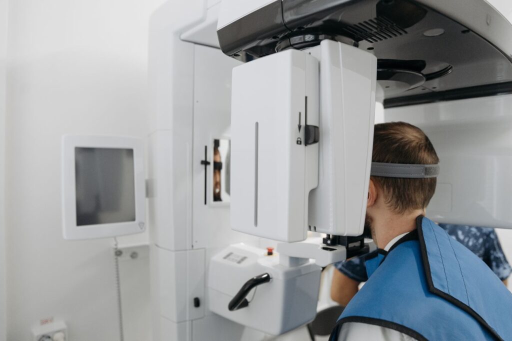
    Undramatic, which is rather the point of mentioning it. You sit or stand with your chin resting on a support while the arm rotates once around your head. Ten to twenty seconds, no enclosed tube, nothing injected, no dye to swallow, and you can be spoken to throughout.

    Better information changes the plan, not just the picture
    Better information changes the plan, not just the picture.

    We review the volume with you where it helps — seeing an extra canal on screen explains a treatment plan faster than any description. Our page on the technology used in the practice covers the rest of the equipment, and the joint position statement on cone-beam imaging in endodontics sets out the professional criteria.

    Frequently asked questions

    Is a 3D scan more radiation than an X-ray?

    Yes, but less than people assume. A limited field-of-view endodontic scan delivers roughly the dose of about two standard periapical radiographs, and it can save taking several conventional images in a complex case.

    Will I need one?

    Probably not. Most root canal treatment is planned from standard radiographs. Scans are taken selectively — for retreatment, suspected extra canals, resorption, trauma, or before surgery.

    What does the scan feel like?

    Nothing. You sit or stand still while the machine rotates around your head for ten to twenty seconds. There is no enclosed tube and no injection.

    Can my dentist see the scan too?

    Yes. Where a scan informs the treatment plan, the findings go into the report we send back to your dentist, along with post-treatment radiographs.

    Have Questions or Ready to Schedule?

    Our team is here to help — call, email, or request an appointment online.
    About this guide. Written for patients of Southeast Endodontics, PC, Charleston, SC. Whether a scan is indicated for your tooth is a clinical judgement made at the consultation, not a routine part of every visit.

  • Tooth Resorption: When a Tooth Dissolves From Within

    Tooth Resorption: When a Tooth Dissolves From Within

    Resorption is one of the few dental conditions where the body dismantles its own tooth structure. It is usually painless, frequently invisible until it is advanced, and almost always found by accident.

    Here is what it is, why an old injury decades ago can still be the cause, and what determines whether a resorbing tooth can be kept.

    Key takeaways

    • Specialised cells break down dentine from either inside the canal or the outer root surface.
    • It is typically painless, so it is usually spotted on a radiograph rather than reported as a symptom.
    • Past trauma is the commonest trigger, sometimes many years earlier.
    • Prognosis depends almost entirely on how early it is found.

    What resorption actually is

    Your body constantly remodels bone using cells that dissolve hard tissue and cells that rebuild it. Teeth are normally protected from this by their outer layers.

    When that protection is damaged — by injury, inflammation or pressure — those dissolving cells can gain access to dentine and start removing it. Nothing rebuilds it, so the process is one-way. In children this is entirely normal and it is how baby teeth loosen; in an adult tooth it is pathological.

    Internal and external resorption

    The main types, and what each means
    TypeWhere it startsTypical causeOutlook
    InternalInside the root canal, working outwardChronic pulp inflammation after trauma or deep decayGood if treated before perforation
    External cervicalAt the neck of the root, under the gumTrauma, orthodontics, older internal bleachingDepends heavily on how far it has spread
    External inflammatoryRoot surface, driven by infectionUsually follows avulsion or luxation injuryNeeds prompt root canal treatment
    External replacementRoot fuses to bone and is replaced by itSevere trauma, often a dried-out replanted toothPoor — the tooth is gradually lost

    Swipe the table sideways to see every column.

    Internal starts in the canal; external starts on the root surface
    Internal starts in the canal and works outward; external starts on the root surface and works in.

    That last row is why how a knocked-out tooth is handled in the first thirty minutes matters so much. A tooth allowed to dry out before replanting frequently develops replacement resorption years later.

    Why it is usually found by accident

    Resorption has almost no early symptoms. It does not usually cause pain, the tooth stays firm, and in external cervical resorption the lesion is hidden beneath the gum.

    A faint pink patch through the crown is the classic visible sign
    A faint pink patch showing through the crown is the classic visible sign.

    The one visible sign, when it appears, is a faint pink area showing through the crown — resorptive tissue is vascular, and its colour shows through thinning enamel. By the time that is visible the lesion is well established.

    Most cases are therefore found on a radiograph taken for another reason, which is a good argument for keeping routine check-ups even when nothing hurts.

    The trauma connection

    The single most common history is an injury to that tooth in the past. A childhood fall, a sports impact, a knock that seemed to resolve — and then twenty years later a radiograph shows resorption.

    An old knock, sometimes decades earlier, is the commonest trigger
    An old knock, sometimes decades earlier, is the commonest trigger.

    That delay is why the connection is so often missed. Orthodontic movement and older internal bleaching techniques are also associated with it, and chronic inflammation around the tooth can contribute. In a meaningful proportion of cases no cause is ever established.

    The signs, such as they are

    Resorption is defined more by the absence of symptoms than the presence of them, which is why the list below is short and mostly late.

    Signs of resorption and what each suggests
    What is noticedWhat it suggestsStage
    Nothing at all — found on a routine filmAny typeEarly, and the best case
    A pink patch showing through the crownInternal, or advanced cervicalEstablished
    A notch or roughness felt at the gumlineExternal cervicalEstablished
    Gum bleeding or overgrowth at one spotExternal cervical with tissue ingrowthEstablished
    Sensitivity or ache in a previously injured toothInflammatory, pulp becoming involvedLater
    Tooth feels solid but sounds dull on tappingReplacement resorptionAdvanced

    Swipe the table sideways to see every column.

    That last one is worth explaining. A tooth fusing to bone loses the slight cushioning of the ligament, so tapping it produces a higher, harder note than its neighbours. It is a small sign with a serious meaning.

    Why 3D imaging changed the diagnosis

    A standard radiograph flattens a three-dimensional root into one image. A resorptive defect on the cheek or tongue side of a root is often hidden behind the root itself.

    Resorption is frequently invisible on a standard film until it is advanced
    Resorption is frequently invisible on a standard film until it is advanced.

    Cone-beam imaging shows the root in cross-section, which reveals both whether resorption is present and how far around the root it has travelled. That second question decides treatment, so a case that looked borderline on a film often becomes clear-cut on a scan — see what a 3D scan shows that an X-ray cannot.

    What treatment involves

    The principle is straightforward: remove the tissue driving the process and seal the defect so it cannot continue.

    Treatment means removing the resorptive tissue and sealing the defect
    Treatment means removing the resorptive tissue and sealing the defect.

    For internal resorption that means root canal treatment, since the process is driven from within the canal. For external cervical resorption, the lesion is usually approached from outside, cleaned out and repaired with a biocompatible material — sometimes with root canal treatment as well, sometimes without if the pulp is uninvolved.

    Extensive lesions that have spread circumferentially around the root, or reached deep below the bone level, are frequently not restorable. That is an honest limitation rather than a failure of technique.

    Monitoring, and why it is worth it

    Not every resorptive lesion needs immediate intervention. Small, stable, symptomless lesions are sometimes monitored with periodic radiographs rather than treated, particularly where treatment would itself be destructive.

    Caught early, many resorbing teeth are kept for years
    Caught early, many resorbing teeth are kept for years.

    What that requires is actually attending the reviews. Resorption is slow but progressive, and the difference between a repairable defect and an unrestorable one is often a couple of years of not being looked at.

    If you have had a significant injury to a front tooth at any point, mention it — it changes what we look for. The American Association of Endodontists’ guide to differentiating resorption covers the classification clinically.

    Frequently asked questions

    What causes tooth resorption?

    Most often a past injury — sometimes decades earlier. Orthodontic movement, internal bleaching using older techniques, and chronic gum inflammation are also associated with it. In a proportion of cases no cause is ever identified.

    Does it hurt?

    Usually not, which is the problem. External cervical resorption in particular is painless and hidden under the gum, so it is often found on a routine radiograph long after it started.

    Can a resorbing tooth be saved?

    Often, if it is caught early. Small, accessible lesions can be cleaned out and repaired. Extensive lesions that have spread around or deep into the root have a poor outlook and may need extraction.

    Is it contagious to other teeth?

    No. It is a localised process affecting one tooth, not an infection that spreads. Having it in one tooth does not mean others will develop it, though a shared cause such as past trauma can affect neighbouring teeth.

    Have Questions or Ready to Schedule?

    Our team is here to help — call, email, or request an appointment online.

    About this guide. Written for patients of Southeast Endodontics, PC, Charleston, SC. Resorption types overlap in presentation and are distinguished by imaging — this is background, not a diagnosis.

  • Gum Problem or Tooth Problem? How to Tell

    Gum Problem or Tooth Problem? How to Tell

    A swelling on the gum beside a tooth has two quite different possible origins, and they need opposite treatments. Getting the distinction right is one of the more consequential calls in dentistry.

    Here is how a gum-origin problem differs from a tooth-origin one, the tests that separate them, and what happens when it turns out to be both.

    Key takeaways

    • A periodontal problem starts outside the tooth; an endodontic one starts inside it.
    • Pulp testing is the single most useful test — a tooth that responds normally to cold is rarely the source.
    • Deep, narrow probing depths point to a periodontal origin; a sudden swelling with a dead pulp points to endodontic.
    • Sometimes both are present, and the order of treatment changes the outcome.

    Two different problems that look alike

    Both produce swelling, tenderness, a bad taste and sometimes pus. What differs is where the infection began.

    A periodontal problem starts in the tissues around the tooth — gum, ligament and supporting bone — usually where a deep pocket has trapped bacteria. The tooth itself may be entirely healthy inside.

    An endodontic problem starts inside the tooth. The pulp dies, infection travels out through the root tip, and finds a route to the surface through the gum. The gum is where you see it, not where it began — our abscess guide covers that in detail.

    How they differ in practice

    Periodontal origin vs. endodontic origin
    FeaturePeriodontalEndodontic
    Pulp response to coldNormal — the tooth is aliveNo response — the pulp is dead
    Probing depthsDeep and wide, often several sitesNormal, or one narrow deep track
    Other teeth affectedFrequently — it is usually generalisedAlmost always a single tooth
    Swelling positionBeside the tooth, nearer the gum marginNearer the root tip, higher in the gum
    Tooth mobilityCommon, from bone lossUnusual unless the infection is extensive
    OnsetGradual, with a history of gum diseaseOften sudden, sometimes after a period of toothache
    History of decay or a big fillingNot necessarilyUsually

    Swipe the table sideways to see every column.

    The tests that settle it

    Pulp testing does most of the work. Cold is applied briefly and we watch how the tooth responds compared with its neighbours.

    A tooth that responds normally to cold is unlikely to be the source
    A tooth that responds normally to cold is rarely the source of the problem.

    A normal response means a living pulp, which makes an endodontic origin unlikely. No response at all means a dead pulp, which makes it very likely. That single test resolves a large majority of cases in under a minute.

    A deep, narrow pocket points to a periodontal origin
    A deep, narrow pocket at one point tells a different story from generalised deep pockets.

    Probing adds the second half. A fine probe is walked around each tooth measuring the gum attachment. Generalised deep pockets across several teeth indicate periodontal disease; a single narrow deep track beside an otherwise healthy gum is usually an endodontic infection draining out.

    What the gum tells you on its own

    Some signs point at the gums before any test. Bleeding when you brush, gums that have receded across the mouth, visible tartar and persistent bad breath are features of periodontal disease rather than of one dying tooth.

    Generalised bleeding and recession point away from a single tooth
    Generalised bleeding and recession point away from any single tooth.

    Conversely, a single tooth that has been aching, is sensitive to hot, or has darkened, with healthy gums elsewhere, is telling you the problem is inside that tooth — see what sensitivity patterns mean and why a tooth darkens.

    When it is both

    Combined lesions exist, and they are the reason this distinction gets its own literature. An endodontic infection draining down the side of a root can create a pocket that then behaves periodontally; advanced periodontal disease reaching the root tip can compromise the pulp.

    Sometimes both are present, and the order of treatment matters
    Combined lesions are less common but change the treatment sequence.

    The practical rule is to treat the endodontic component first. A tooth with a dead pulp will not respond to periodontal treatment while the source is untreated, whereas resolving the pulp infection frequently allows the associated pocket to heal on its own. Reversing that order wastes months.

    Why the distinction matters so much

    The treatments have almost nothing in common. Periodontal disease is managed by cleaning below the gum, sometimes surgically, plus daily control at home — a long-term maintenance problem.

    Treating the wrong one wastes months and can cost the tooth
    Treating the wrong one wastes months, and occasionally costs the tooth.

    An endodontic infection is resolved by treating the inside of the tooth once. No amount of scaling reaches a dead pulp, and no amount of root canal treatment fixes a deep periodontal pocket.

    Prognosis differs too. A tooth with an endodontic problem and healthy support has an excellent outlook. A tooth that has already lost most of its bone support has a poor one regardless of what is done inside it — which is one of the situations where extraction is the honest recommendation.

    What each one actually needs

    Treatment, prognosis and who provides it
    Periodontal originEndodontic origin
    TreatmentCleaning below the gum, sometimes surgicallyRoot canal treatment
    Number of visitsA course, then ongoing maintenanceUsually one, occasionally two
    Your role afterwardsDaily control is decisiveNormal brushing; nothing special
    Who provides itGeneral dentist or periodontistEndodontist or general dentist
    OutlookDepends on remaining bone supportGood where support is intact
    If left untreatedProgressive bone loss, eventual mobilityAbscess, then spreading infection

    Swipe the table sideways to see every column.

    The column that surprises people is the third row. Periodontal disease is controlled rather than cured, and how well it is controlled depends largely on what happens at home between appointments. An endodontic infection is dealt with once and does not come back if the tooth is properly restored.

    What to do if you have a swelling now

    Get it looked at rather than self-diagnosing. Both origins are treatable and both worsen if left, and the tests take minutes.

    Periodontal problems respond to daily control; endodontic ones do not
    Periodontal problems respond to daily control; endodontic ones do not.

    Go the same day if the swelling is spreading, you have a fever, or the area near your eye or throat is involved — that is covered in the emergency guide and applies whichever the origin. The American Academy of Periodontology’s information on gum disease covers the periodontal side in more depth.

    Frequently asked questions

    Can a gum infection kill the nerve in a tooth?

    It can, though it is the less common direction. Severe periodontal disease reaching the root tip can affect the pulp through the small openings there. The reverse — an infected pulp draining out through the gum — is seen far more often.

    Why does the swelling keep coming back after antibiotics?

    Because antibiotics treat the infection but not its source. If the source is a dead pulp, the tooth needs root canal treatment; if it is a deep periodontal pocket, that pocket needs cleaning. Either way the swelling returns once the course ends.

    Does it matter which one it is if both need treating?

    Very much. The order changes the outcome. Where the pulp is the origin, treating it first often lets the gum problem resolve on its own; treating the gum first while the source is untreated achieves little.

    Will an X-ray show which it is?

    It helps but rarely decides it. Bone loss patterns differ, but early changes are subtle and both conditions can produce similar shadows. Pulp testing and probing depths carry more weight than the film.

    Have Questions or Ready to Schedule?

    Our team is here to help — call, email, or request an appointment online.

    About this guide. Written for patients of Southeast Endodontics, PC, Charleston, SC. We diagnose and treat the endodontic side; periodontal treatment is provided by your general dentist or a periodontist, and combined cases need both.

  • Why Won’t My Tooth Get Numb?

    Why Won’t My Tooth Get Numb?

    Few things undermine confidence faster than feeling something you were told you would not. If a tooth has refused to go numb before, you are not imagining it.

    There are well-documented reasons why some teeth resist anaesthetic, and established techniques for dealing with them. Here is what is actually happening, and what should be done about it.

    Key takeaways

    • An acutely inflamed pulp is chemically harder to anaesthetise — this is a property of the tooth, not of you.
    • Standard lower-jaw blocks succeed in 85–90% of healthy teeth but can drop below 30% in a “hot” tooth.
    • Repeating the same failed injection rarely helps; changing technique does.
    • Tell us the moment you feel something sharp. That is information, not an interruption.

    Why an inflamed tooth resists anaesthetic

    Local anaesthetic works by blocking sodium channels in nerve fibres, which stops the pain signal reaching your brain. In a healthy tooth that is straightforward.

    An acutely inflamed pulp resists the standard block more than any other tooth
    The problem is the state of the pulp, not the amount of anaesthetic used.

    An acutely inflamed pulp is a different environment. The inflamed tissue is more acidic, which reduces how much of the drug crosses into the nerve. The nerve fibres themselves become sensitised and fire at lower thresholds, and inflammation upregulates channel types that ordinary anaesthetic blocks less effectively.

    Clinically this is called a “hot” tooth, and the numbers are striking. Standard inferior alveolar nerve blocks achieve profound pulpal anaesthesia in roughly 85–90% of uninflamed teeth. In symptomatic irreversible pulpitis, published success for the same block can fall below 30%.

    Why lower back teeth are hardest

    If you have had trouble, it was probably a lower molar. Upper teeth are anaesthetised by depositing solution near the root, through relatively porous bone. That is reliable.

    Lower molars are the hardest teeth in the mouth to anaesthetise reliably
    Lower molars are the hardest teeth in the mouth to anaesthetise reliably.

    Lower back teeth sit in dense bone anaesthetic will not penetrate, so the nerve must be blocked further back, before it enters the jaw. That target is small and its position varies between people.

    Why some teeth are harder than others
    FactorEffect on anaesthesia
    Acutely inflamed pulpLargest single factor — block success can fall below 30%
    Lower molarDense bone; the nerve must be blocked further back
    Anatomical variationThe nerve is not where the textbook says in a minority of people
    Accessory innervationA second nerve supply the main block does not reach
    Existing infectionAcidic tissue reduces drug uptake
    AnxietyLowers pain threshold; sharpens what is felt

    Swipe the table sideways to see every column.

    What should happen when the first injection fails

    The wrong response is more of the same. If a standard block has not worked, repeating it usually will not either, and there are safe limits on total dose.

    Supplemental techniques deliver anaesthetic much closer to the nerve
    Supplemental techniques deliver anaesthetic far closer to the nerve.

    The right response is a different technique. Several are well established and routine in specialist practice:

    • Intraosseous injection. Anaesthetic is placed directly into the bone beside the tooth. Onset is 10–20 seconds and it is the most predictable option for a hot tooth.
    • Intraligamentary injection. Delivered into the ligament space around the root; reported success of 50–96% as a supplemental technique.
    • Intrapulpal injection. Directly into the pulp once accessed — briefly uncomfortable, then immediately effective.
    • An alternative block. Gow-Gates and Vazirani-Akinosi techniques target the nerve higher up, bypassing the anatomical variation that defeats the standard approach.

    None are exotic. They are routine in a practice treating inflamed pulps daily — part of why “we could not get you numb” is a recognised reason to refer.

    What you should do

    Speak up immediately. Patients often endure sharp sensations out of politeness. It is not meant to feel like that, and enduring it makes the next appointment harder.

    A raised hand is information we need, not an interruption
    A raised hand is information we need, not an interruption.
    1. Tell us at the start if you have had trouble getting numb before. It changes the plan from the first injection rather than the third.
    2. Agree a stop signal — a raised hand pauses everything immediately.
    3. Describe what you feel. Pressure and vibration are expected; sharp, hot or electric sensations are not. That distinction tells us a great deal.
    4. Do not tough it out. There is no prize, and it works against you.

    What a supplemental injection feels like

    People understandably worry that a second, different injection means something has gone wrong. It does not — it means the first technique met a tooth it was never going to beat.

    The supplemental techniques, and what to expect from each
    TechniqueWhat you feelHow fast it works
    IntraosseousBrief pressure; sometimes a fast heartbeat for a minute10–20 seconds
    IntraligamentaryFirm pressure beside the toothUnder a minute
    IntrapulpalA few seconds of sharp sensation, then nothingImmediate
    Gow-Gates blockMuch like a standard block, placed higher5–10 minutes

    Swipe the table sideways to see every column.

    The brief racing heartbeat some people notice after an intraosseous injection is adrenaline reaching the bloodstream faster. It is harmless and passes within a minute.

    Anxiety, and why it is not “just in your head”

    Anxiety does not stop anaesthetic working, but it changes how a partially anaesthetised tooth is experienced. A heightened nervous system registers pressure as pain and mild sensation as sharp.

    Anxiety genuinely lowers pain threshold, which compounds the problem
    Anxiety genuinely lowers pain threshold, which compounds the problem.

    It also creates a loop: a difficult appointment increases anxiety, which makes the next one harder. Breaking that loop is worth doing deliberately — our guide to sedation options covers both the medication and the non-drug approaches, and sedation combined with proper technique handles nearly all of these cases.

    Why getting there matters clinically

    This is not only about comfort. An inadequately anaesthetised tooth is difficult to treat properly, because the patient cannot stay still and the clinician is working against the clock.

    Almost every tooth can be made comfortable with the right technique
    Almost every tooth can be made comfortable with the right technique.

    Rushed treatment is how canals get missed, which is a leading route to treatment failing later. Taking the extra ten minutes to achieve profound anaesthesia is part of doing the job well, not a delay to it.

    It also argues against waiting. The more acutely inflamed a tooth becomes, the harder it is to numb — so a tooth treated early is easier to make comfortable than the same tooth treated three weeks later. If symptoms are escalating, the emergency guide covers what should not wait.

    If a previous appointment went badly, say so when you book — see what a first visit involves. The published trial of management after inferior alveolar nerve block failure covers the clinical evidence behind the techniques above.

    Frequently asked questions

    Why did the injection work last time but not now?

    Because the tooth is different, not you. An acutely inflamed pulp changes chemically and electrically in ways that make it resist local anaesthetic. The same injection in the same jaw on a healthy tooth usually works perfectly.

    Does being anxious make it harder to get numb?

    Indirectly, yes. Anxiety lowers pain threshold and makes sensations register as sharper, so a partially anaesthetised tooth feels worse than it otherwise would. Managing the anxiety genuinely helps.

    Will more anaesthetic eventually work?

    Not necessarily — repeating the same injection that has already failed rarely succeeds, and there are safe limits. Changing technique works far better than increasing volume.

    Is it true that redheads need more anaesthetic?

    There is some published evidence of altered anaesthetic requirements associated with certain genetic variants. It is worth mentioning, but it is a minor factor next to whether the pulp is acutely inflamed.

    Have Questions or Ready to Schedule?

    Our team is here to help — call, email, or request an appointment online.
    About this guide. Written for patients of Southeast Endodontics, PC, Charleston, SC. Success figures are published averages for the techniques described; individual anatomy varies more than most people expect.