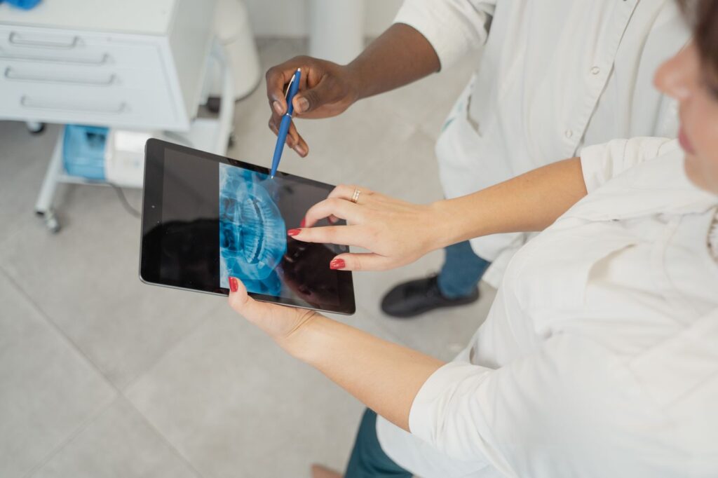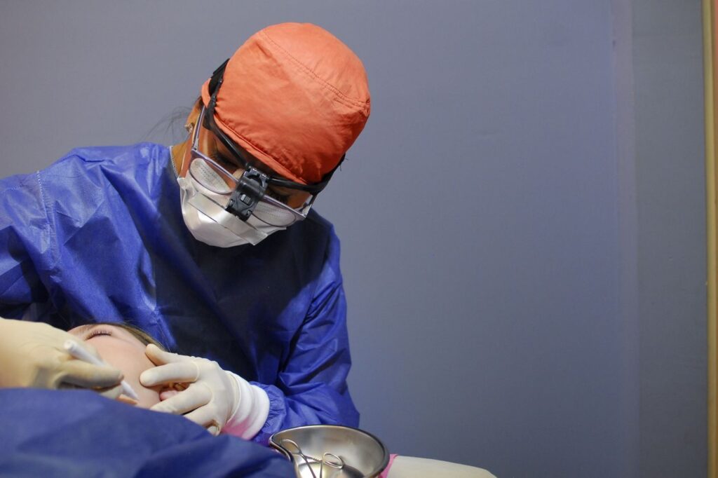
When a root canal has been done well and the tooth still will not settle, the problem is usually at the very tip of the root — the one part that cannot always be reached from inside the tooth.
An apicoectomy solves that by approaching from the outside instead. Here is what the procedure involves, what recovery is actually like, and when it is the right answer rather than retreatment or extraction.
Key takeaways
- The root tip and surrounding infected tissue are removed through the gum, and the root end is sealed.
- It takes 30–90 minutes and is done under local anaesthetic.
- Microsurgical technique reports around 90% success or better — far above older published figures.
- It is a second-line option: non-surgical retreatment is normally tried first.
Why the root tip is the problem
A root canal cleans and seals the canal from the crown downwards. At the very end of the root, the canal frequently stops being a single tube and becomes a fine delta of branches too narrow for any instrument to enter.
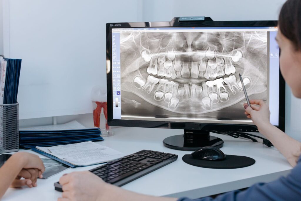
Bacteria surviving in that mesh keep the surrounding bone inflamed. No amount of cleaning from inside reaches them, which is why a technically excellent root canal can still leave a tooth that aches on biting or shows a persistent shadow on a radiograph.
When it is the right choice
Apicoectomy is not the first response to a failing root canal. Non-surgical retreatment is generally tried first, because it addresses the whole canal rather than just the end of it.
| Situation | Why retreatment is unsuitable |
|---|---|
| A well-fitting post and crown | Removing them risks fracturing the root or destroying a sound restoration |
| A separated instrument blocking the canal | The blockage cannot be bypassed from inside |
| Retreatment already attempted and failed | The canal has been cleaned as far as it can be |
| A cyst or lesion needing biopsy | Tissue can only be removed and examined surgically |
| A perforation low in the root | Repair from outside is more predictable |
Swipe the table sideways to see every column.
What actually happens
- Anaesthetic. The area is numbed thoroughly, including the surrounding bone.
- Access. A small incision is made in the gum beside the tooth and the tissue lifted to expose the bone over the root tip.
- Removal. A few millimetres of the root tip are removed, along with the infected tissue around it.
- Sealing. The cut end of the root is prepared and sealed with a biocompatible filling material.
- Closure. The gum is repositioned and closed with fine sutures, usually removed after a week.
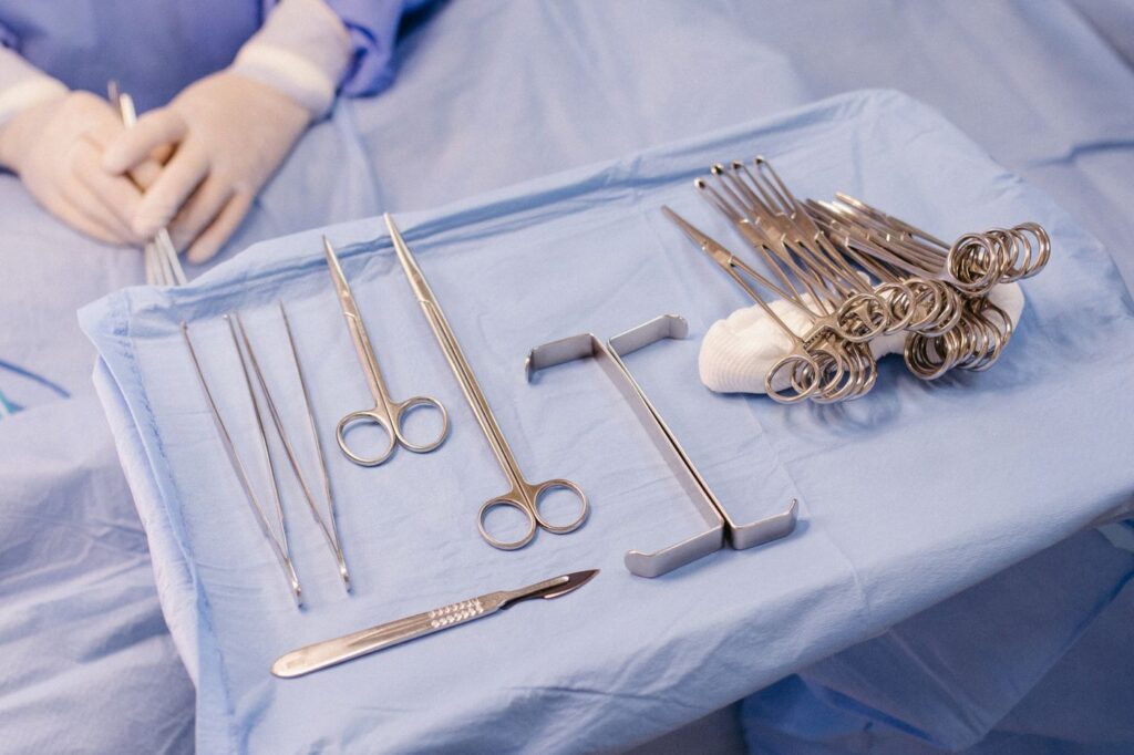
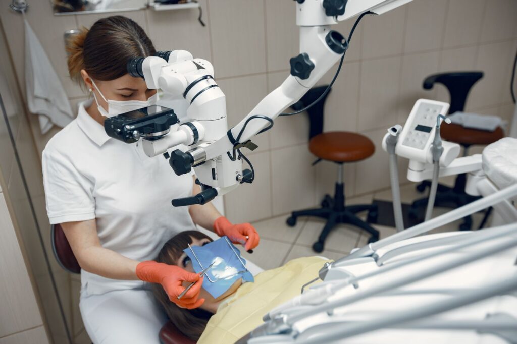
Magnification is the single biggest change in this procedure over the last twenty years. Working under a microscope allows a much smaller opening, a cleaner cut and a far better seal — see the equipment we use.
Recovery, honestly
Expect soreness and swelling. It peaks at about 48 hours and then improves daily, and a cold compress in the first day genuinely helps.

| When | What to expect | What to do |
|---|---|---|
| Day of surgery | Numbness, then soreness as it wears off | Cold compress, anti-inflammatories, rest |
| Day 1–2 | Swelling peaks; possible bruising | Soft food, no vigorous rinsing or brushing at the site |
| Day 3–7 | Steady improvement; sutures removed around day 7 | Return to normal activity; gentle cleaning |
| Week 2–4 | Soft tissue fully healed | Normal brushing and diet |
| Month 3–6 | Bone fills in at the root tip | Follow-up radiograph to confirm healing |
Swipe the table sideways to see every column.
Most patients return to work the following day. What takes longer is invisible: the bone defect left by the infection fills in over three to six months, which is why we review with a radiograph rather than judging by symptoms.
What the success rate really means
You will find figures ranging from 60% to 97% for this procedure, and the spread is not noise — it is chronology.
Older studies describe surgery done without magnification, with materials long superseded. Contemporary microsurgical series report around 90% or better, with roughly 97% still successful at five years and more than 75% at ten to thirteen years. If you are reading an alarming number, check when the study was published.
What it costs
An apicoectomy typically runs somewhere between a molar root canal and an extraction with an implant. It is billed as a surgical endodontic procedure, which most plans cover at the same percentage as other endodontic work — though it draws on the same annual maximum.
Ask for a written estimate with your benefits checked before treatment. Our cost guide explains how deductibles and annual maximums interact, and why timing across two plan years sometimes helps.
The risks worth knowing about
It is minor surgery, and minor surgery is not no surgery. Bruising and swelling are expected rather than complications.
The specific risks depend on where the tooth sits. Upper back teeth are close to the maxillary sinus, and lower back teeth to the nerve supplying the lip and chin, so we assess both on 3D imaging before operating rather than discovering the relationship during the procedure. Numbness of the lip is uncommon and usually temporary where it occurs.
The other honest caveat is that not every case is suitable. A tooth with a vertical root fracture, or too little bone support remaining, will not be improved by surgery — and we would tell you that rather than attempt it.
Weighing it against extraction
The honest comparison is not surgery versus doing nothing, but surgery versus removing the tooth and replacing it.

An apicoectomy keeps your own root, ligament and bone, and costs considerably less than extraction plus an implant. It is also reversible in the sense that extraction remains available afterwards — the reverse is not true. The alternatives compared on ten-year cost sets out the numbers.
Our apicoectomy procedure page covers the clinical detail, and endodontic surgery the wider category. The published analysis of outcomes after non-surgical treatment fails sets surgery in context against the alternatives.
Frequently asked questions
Is an apicoectomy painful?
During, no more than a root canal — the area is fully anaesthetised. Afterwards expect soreness and swelling for a few days, managed with anti-inflammatories and a cold compress. Most people describe it as comparable to having a tooth out, without losing the tooth.
How long does it take?
Between 30 and 90 minutes, depending on which tooth and how accessible the root tip is. Front teeth are usually at the shorter end; lower molars at the longer end.
What is the success rate?
Reported success with modern microsurgical technique is around 90% or better, with roughly 97% still doing well at five years in some series. Older figures quoting 60–70% predate the microscope and current materials.
Can it be repeated if it fails?
Sometimes, but a second surgery on the same root has a lower success rate. If an apicoectomy fails, extraction and replacement is usually the more sensible next step than a repeat attempt.
Have Questions or Ready to Schedule?
Our team is here to help — call, email, or request an appointment online.