10-E Farmfield Avenue, Charleston, SC 29407

235-213-0595

Category: Root Canals

Guides to root canal treatment, alternatives, and what to expect from endodontic care.

  • Root Canals in Older Adults: What Changes and What Doesn’t

    Root Canals in Older Adults: What Changes and What Doesn’t

    People in their seventies and eighties routinely ask whether a root canal is worth it at their age. The evidence answers that clearly: age does not affect whether treatment works.

    What does change is how the tooth behaves and what else is going on medically. Here is what genuinely differs about endodontic treatment later in life.

    Key takeaways

    • Across two dozen studies, age is not a prognostic factor for root canal outcome.
    • Canals narrow throughout life, which makes treatment technically harder rather than less successful.
    • Medications matter far more than age in planning treatment.
    • Dry mouth from medication is the commonest driver of new decay later in life.

    What actually changes with age

    Teeth lay down dentine continuously. Every year the pulp chamber gets a little smaller and the canals a little narrower, and by seventy a canal that was straightforward at twenty can be extremely fine.
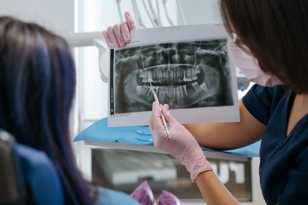
    Canals narrow over a lifetime as secondary dentine is laid down
    Canals narrow over a lifetime as secondary dentine is laid down.

    The pulp itself changes too: more fibrous tissue, less blood supply, fewer nerve fibres. That has a practical consequence — an older tooth often responds more weakly to cold testing, and a reduced response is not necessarily a dead pulp. It has to be read against the neighbouring teeth rather than in isolation.

    Why calcified canals are harder, not hopeless

    A calcified canal is not blocked. It is narrow, and the entrance is often hidden under a layer of dentine laid down over decades.

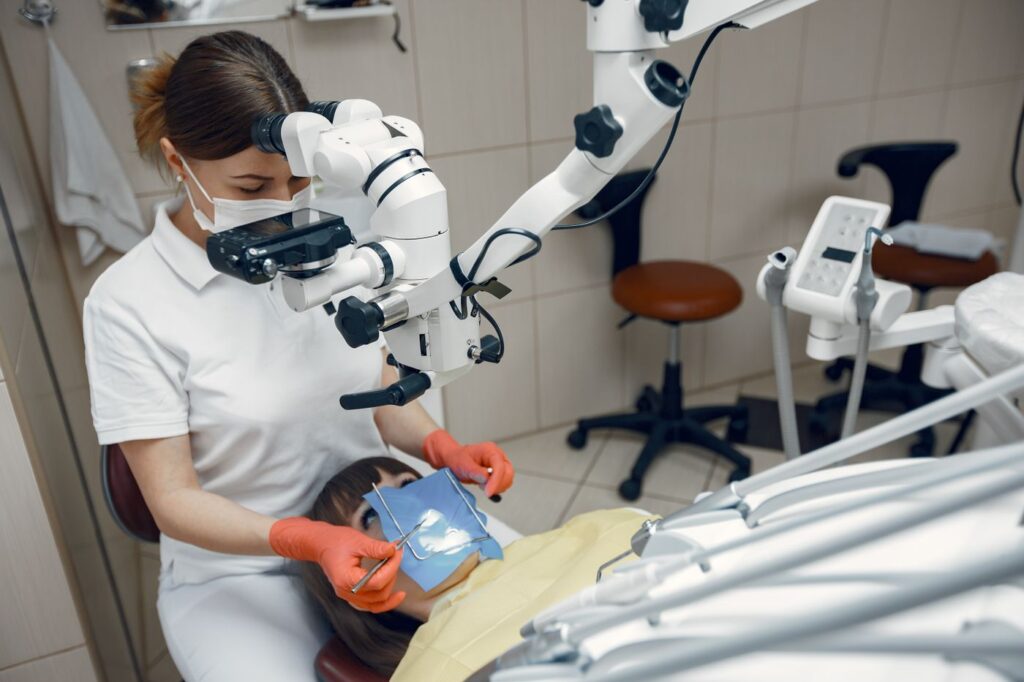
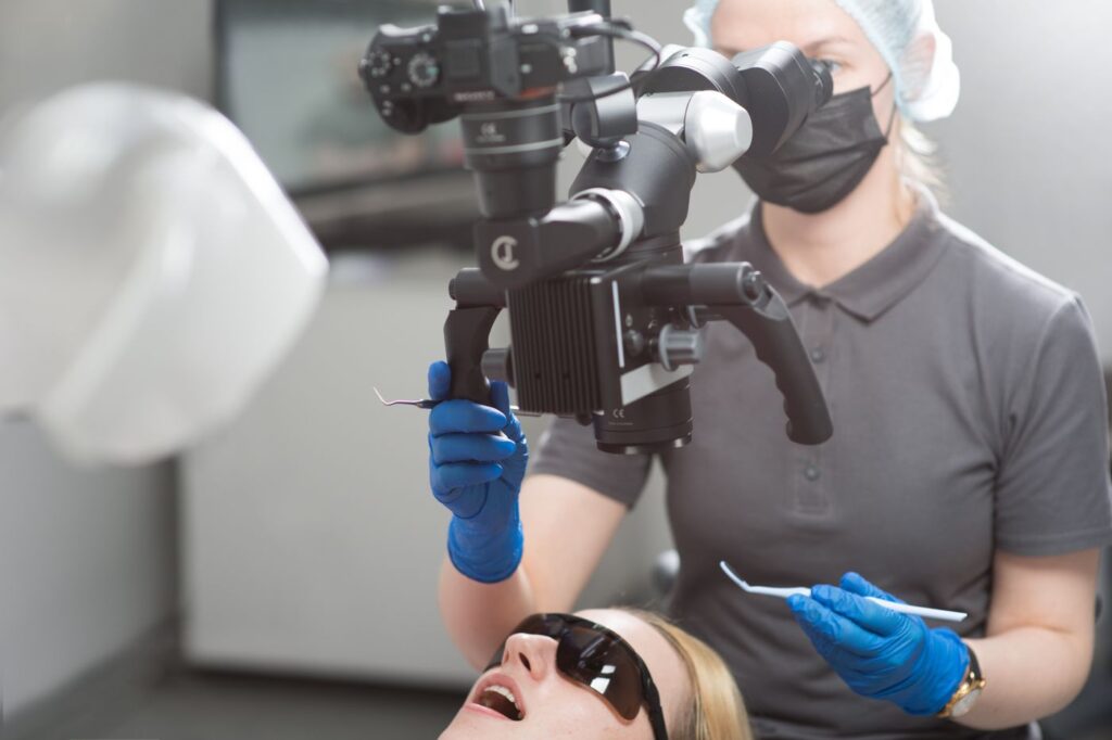
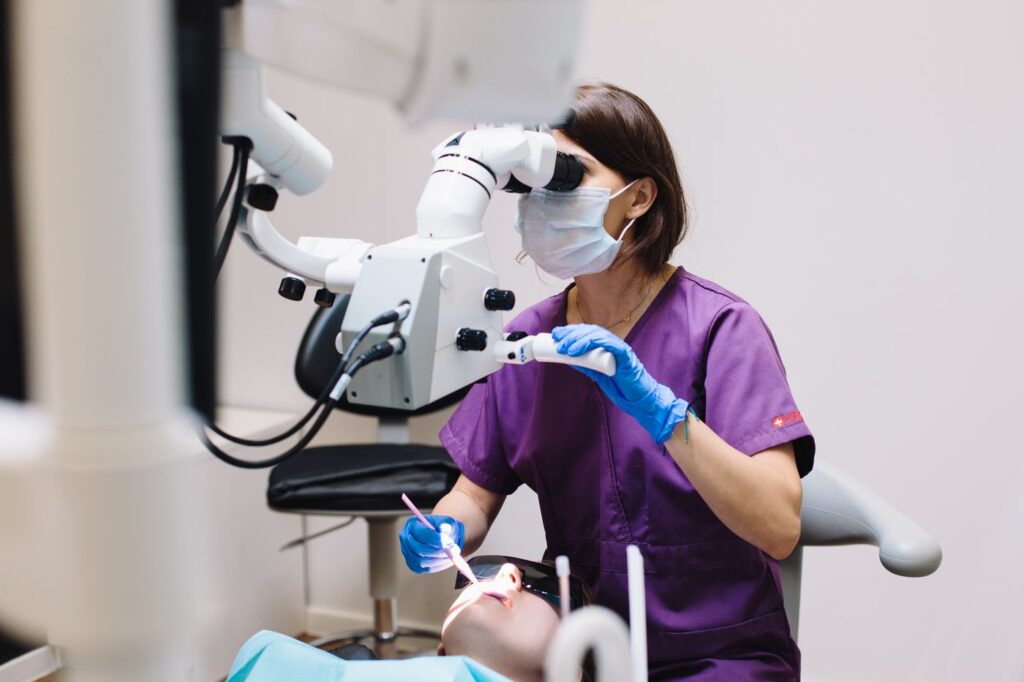
    Finding a calcified canal is a magnification problem more than anything
    Finding a calcified canal is largely a magnification problem.

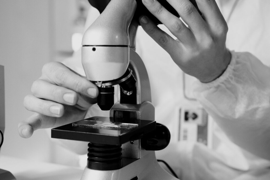
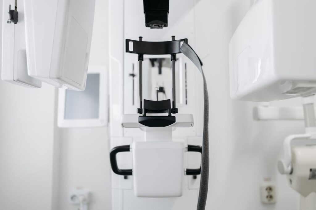
    Locating it is a magnification and imaging problem more than a mechanical one. Under a microscope the subtle colour change marking a canal entrance is visible; without one it frequently is not. 3D imaging shows whether a canal exists and where it runs before any drilling starts, which matters when the alternative is exploring blind.

    Appointments take longer as a result. That is worth knowing in advance rather than being surprised by it, and it is one of the clearer reasons a general dentist may prefer to refer a heavily calcified tooth rather than attempt it — see what usually prompts a referral. The tooth is not more likely to fail; it simply needs equipment and time that a routine appointment does not allow for.

    What matters more than age

    Factors that genuinely affect planning
    FactorWhy it mattersWhat we do
    AnticoagulantsBleeding during surgical proceduresUsually continued; coordinated with your physician
    Bisphosphonates or denosumabAffect bone healing after extractionOften makes saving the tooth preferable to removing it
    Dry mouth from medicationSharply raises decay riskPrevention advice, and expect more frequent review
    DiabetesSlower periapical healingLonger follow-up before judging success
    Difficulty lying backComfort and breathingShorter appointments, adjusted chair position
    Limited mouth openingAccess to back teethPlanned in advance rather than discovered

    Swipe the table sideways to see every column.

    Medication lists matter more than age in planning treatment
    Medication lists matter more than age in planning treatment.

    The bisphosphonate row is the one people underestimate. If you take or have taken these drugs, extraction carries a small but real risk to the jawbone — which shifts the balance firmly toward keeping the tooth. That reverses the usual assumption that pulling is the simpler option.

    Comfort during a longer appointment

    If the technical work takes longer, the practical question becomes whether you can comfortably stay in the chair for it. That is worth raising at the consultation rather than enduring on the day.

    Difficulty lying flat is the commonest issue, whether from breathing, reflux or a bad back. The chair position can be adjusted, and treatment can be divided across two shorter visits instead of one long one. Neither compromises the result.

    Anaesthetic works no differently with age, though it can take slightly longer to wear off. If you take medication for blood pressure or heart rhythm, tell us — it occasionally changes which anaesthetic we choose, and it is easier to plan for than to discover. Sedation options remain available and are assessed against your medical history rather than ruled out by age.

    Decades of previous dentistry

    Older teeth rarely arrive untouched. Crowns, posts, large fillings and previous root canals all change the approach.
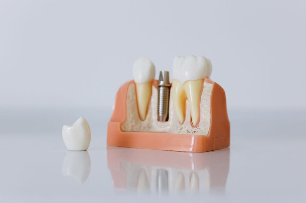
    Decades of previous dentistry change how a tooth is accessed
    Decades of previous dentistry change how a tooth is accessed.

    Reaching the pulp through an existing crown means cutting through it, and the crown may or may not survive. Where a post is present, removing it carries a risk of fracturing the root — which is one of the situations where root-end surgery is preferred over conventional retreatment.

    Dry mouth, and why it causes so much trouble

    Hundreds of common medications reduce saliva — blood pressure drugs, antidepressants, antihistamines, diuretics. Saliva buffers acid, washes away debris and carries minerals that repair early damage, so losing it changes the mouth substantially.

    Dry mouth from medication is the commonest driver of decay later in life
    Dry mouth from medication is the commonest driver of decay later in life.

    The characteristic result is decay at the gumline and around the edges of old restorations, in people who went decades without a cavity. It is worth raising with whoever prescribes the medication and worth managing actively, because it is the commonest route to needing a root canal at seventy.

    Is it worth treating?

    This is the question behind the question, and it deserves a direct answer. Success rates are the same; a treated tooth lasts as long as any other; and the alternatives have their own costs.

    Keeping natural teeth into later life is now the expectation, not the exception
    Keeping natural teeth into later life is now the expectation, not the exception.

    Extraction means either a gap that affects chewing, or a replacement. Implants take months and need adequate bone; dentures change what you can eat. The alternatives compared covers the trade-offs, and the calculation frequently favours keeping the tooth more strongly at seventy than at forty.

    Saving the tooth vs. replacing it, later in life
    ConsiderationRoot canal and crownExtraction and implant
    Time to a finished resultWeeksSeveral months
    Number of appointmentsTypically two to threeConsiderably more
    Depends on bone volumeNoYes — may need grafting first
    Affected by bisphosphonate historyNoYes, and significantly
    Surgical procedure requiredUsually noneYes
    Keeps the natural root and ligamentYesNo

    Swipe the table sideways to see every column.

    The time column is the one that decides it for many people. Months of a gap, a temporary and repeated visits is a real cost when the alternative finishes in a fortnight.

    Published work on endodontic considerations in older patients covers the clinical detail behind this.

    Frequently asked questions

    Am I too old for a root canal?

    No. Across two dozen studies, patient age does not affect the outcome of root canal treatment. The technical demands can be higher, but the results are not worse.

    Why does my dentist say the canals are calcified?

    Because they narrow throughout life. Teeth lay down dentine continuously, so a canal that was wide at twenty can be very fine at seventy. It makes the tooth harder to treat, not impossible.

    I take several medications. Does that matter?

    It matters more than your age does. Anticoagulants, bisphosphonates and drugs causing dry mouth all affect planning, so bring a current list to the appointment.

    Is extraction simpler at my age?

    Not necessarily. Older bone is denser and roots can be more brittle, so extraction is not automatically the easier option — and replacing the tooth afterwards has its own considerations.

    Have Questions or Ready to Schedule?

    Our team is here to help — call, email, or request an appointment online.
    About this guide. Written for patients of Southeast Endodontics, PC, Charleston, SC. Bring a current medication list and your physician’s details to any appointment — they change planning more than anything else here.

  • Can the Nerve Be Saved? Vital Pulp Therapy in Adults

    Can the Nerve Be Saved? Vital Pulp Therapy in Adults

    For decades, a pulp inflamed past a certain point meant one thing: remove all of it. That position has genuinely shifted, and it is one of the more interesting changes in endodontics.

    Vital pulp therapy keeps living pulp tissue in the tooth rather than removing it entirely. Here is what it involves, the evidence behind it, and the cases where a conventional root canal remains the right answer.

    Key takeaways

    • Only the inflamed part of the pulp is removed; healthy tissue in the roots stays alive.
    • Trials in mature teeth report success comparable with root canal treatment — around 93% in several studies.
    • Less post-operative pain on day one, shorter appointments and lower cost.
    • Candidacy is decided during the appointment, based on how the pulp actually behaves.

    What it actually is

    A conventional root canal removes the pulp from the crown and every root, then fills the space. Vital pulp therapy removes only the part that is inflamed — usually the portion in the crown — and seals a medicated material over the healthy tissue beneath.

    A pulpotomy removes the inflamed crown portion and leaves the roots
    Only the inflamed portion is removed; the rest of the pulp stays alive.

    The reasoning is that pulp inflammation is often localised rather than uniform. The tissue nearest the decay may be beyond saving while the tissue deeper in the roots is perfectly healthy, and removing all of it treats the whole pulp for a problem confined to part of it.

    Why this is newer than it sounds

    Partial pulp treatment is not a new idea. What changed is the materials.

    Modern bioceramic materials are what made this predictable
    Modern bioceramic materials are what made the approach predictable.

    Older capping materials sealed poorly and irritated the tissue they were meant to protect, so results were unreliable and the approach fell out of favour in adults. Hydraulic calcium silicate cements seal well, tolerate moisture, and actively stimulate the pulp to lay down a hard tissue barrier of its own. That is the difference between an optimistic technique and a predictable one.

    What the evidence shows

    Vital pulp therapy vs. root canal treatment in mature teeth
    MeasureFull pulpotomyRoot canal treatment
    Clinical success~97%~99%
    Radiographic success~93%~95%
    Pain on day one after treatmentSignificantly lowerHigher
    Treatment timeShorterLonger
    CostLowerHigher
    Patient satisfactionHigher across time, pain and costLower on those measures

    Swipe the table sideways to see every column.

    Those figures come from randomised trials comparing the two in mature teeth with symptomatic irreversible pulpitis — the exact diagnosis that used to mean an automatic root canal. Longer-term series using calcium silicate materials report success between roughly 78% and 90% at one to five years.

    The honest caveat is that the follow-up periods are shorter than for conventional treatment, which has decades of data behind it. This is well-supported rather than fully settled.

    Who it suits

    The typical case is a tooth where decay has reached the pulp and there are symptoms, but the tooth is otherwise sound and restorable.

    It applies where decay has reached the pulp but the tissue can still recover
    It applies where decay has reached the pulp but the tissue can still recover.

    Candidacy is genuinely decided in the chair. Once the decay is removed and the pulp exposed, what matters is how it bleeds: healthy pulp bleeds bright red and stops within a few minutes under gentle pressure. Bleeding that will not stop indicates inflammation deeper than the part being removed, and treatment converts to a conventional root canal in the same appointment.

    When each approach applies
    SituationUsual approach
    Decay reaching the pulp, bleeding controls quicklyVital pulp therapy
    Bleeding will not stop after several minutesConvert to root canal treatment
    Pulp already dead — no response to coldRoot canal treatment
    Swelling or an abscess presentRoot canal treatment
    Tooth needs a crown for structural reasons anywayOften root canal treatment
    Immature tooth in a child or teenagerVital pulp therapy strongly preferred

    Swipe the table sideways to see every column.

    That last row is long-established practice rather than a new development — see endodontics in children, where keeping the pulp alive lets the root finish forming.

    Why keeping the pulp is worth something

    A tooth with a living pulp retains sensation, which means it can still tell you when something is wrong. A root-filled tooth cannot, so problems there are found on radiographs rather than reported as symptoms.

    A living pulp keeps sensing and defending the tooth
    A living pulp keeps sensing and defending the tooth.

    Living pulp also continues laying down dentine in response to wear and irritation, and less tooth structure is removed in the first place — which matters for long-term fracture resistance, as the survival data on cuspal coverage shows.

    What the appointment is like

    It starts the same way as any other treatment: local anaesthetic, a rubber dam, and the decay removed completely. The difference comes at the point the pulp is reached.

    Instead of proceeding into the roots, the exposed tissue is removed to a defined level and bleeding is controlled under light pressure. That pause is the diagnostic step — what happens in those few minutes decides the rest of the appointment.

    If bleeding settles, the material is placed directly against the remaining pulp and sealed, and the tooth is restored in the same visit. Post-operative discomfort is typically milder than after conventional treatment.

    What it does not change

    The restoration afterwards is as important as it is following a root canal. A well-treated pulp under a leaking filling will fail, because bacteria simply return.

    Shorter appointment, lower cost, and less post-operative pain on day one
    Shorter appointment and lower cost, but the restoration still matters.

    Follow-up matters too. These teeth are reviewed for symptoms and for continued pulp response, and a proportion do go on to need conventional treatment later — which is a manageable outcome rather than a disaster.

    Asking about it

    It is a reasonable question to raise if you have been told you need a root canal on a tooth that is otherwise in good condition. The answer may well be no — but the reasoning is worth hearing.

    Where the pulp is too far gone, conventional treatment remains the answer
    Where the pulp is too far gone, conventional treatment remains the answer.

    What it is not is a way to avoid treatment. A tooth with a dead pulp or an abscess needs conventional root canal treatment, and delaying in the hope of a lesser option makes that worse. The published randomised trial comparing full pulpotomy with root canal therapy is the source for the figures above.

    Frequently asked questions

    Is this the same as the pulp capping done on children?

    The principle is the same — keep living pulp alive — but the evidence in adults is newer. What changed is the materials: hydraulic calcium silicate cements seal and stimulate repair far better than the older options, which is why adult cases now succeed at rates comparable with root canal treatment.

    How would I know if I am a candidate?

    It is decided at the appointment, not before. The deciding factor is what the pulp looks like once the decay is removed and how it bleeds. If bleeding stops within a few minutes under pressure, the tissue is usually healthy enough.

    What happens if it does not work?

    You have a conventional root canal, which was the alternative anyway. Failure means treatment moves on rather than the tooth being lost, and that is the reason it is reasonable to attempt.

    Is it cheaper?

    Usually, yes — published trials report lower cost and shorter treatment time than root canal therapy. It still needs a proper restoration afterwards, which is the larger part of the bill on a back tooth.

    Have Questions or Ready to Schedule?

    Our team is here to help — call, email, or request an appointment online.

    About this guide. Written for patients of Southeast Endodontics, PC, Charleston, SC. Vital pulp therapy is well supported but has shorter follow-up data than conventional treatment; whether it suits your tooth is decided clinically.

  • Tooth Resorption: When a Tooth Dissolves From Within

    Tooth Resorption: When a Tooth Dissolves From Within

    Resorption is one of the few dental conditions where the body dismantles its own tooth structure. It is usually painless, frequently invisible until it is advanced, and almost always found by accident.

    Here is what it is, why an old injury decades ago can still be the cause, and what determines whether a resorbing tooth can be kept.

    Key takeaways

    • Specialised cells break down dentine from either inside the canal or the outer root surface.
    • It is typically painless, so it is usually spotted on a radiograph rather than reported as a symptom.
    • Past trauma is the commonest trigger, sometimes many years earlier.
    • Prognosis depends almost entirely on how early it is found.

    What resorption actually is

    Your body constantly remodels bone using cells that dissolve hard tissue and cells that rebuild it. Teeth are normally protected from this by their outer layers.

    When that protection is damaged — by injury, inflammation or pressure — those dissolving cells can gain access to dentine and start removing it. Nothing rebuilds it, so the process is one-way. In children this is entirely normal and it is how baby teeth loosen; in an adult tooth it is pathological.

    Internal and external resorption

    The main types, and what each means
    TypeWhere it startsTypical causeOutlook
    InternalInside the root canal, working outwardChronic pulp inflammation after trauma or deep decayGood if treated before perforation
    External cervicalAt the neck of the root, under the gumTrauma, orthodontics, older internal bleachingDepends heavily on how far it has spread
    External inflammatoryRoot surface, driven by infectionUsually follows avulsion or luxation injuryNeeds prompt root canal treatment
    External replacementRoot fuses to bone and is replaced by itSevere trauma, often a dried-out replanted toothPoor — the tooth is gradually lost

    Swipe the table sideways to see every column.

    Internal starts in the canal; external starts on the root surface
    Internal starts in the canal and works outward; external starts on the root surface and works in.

    That last row is why how a knocked-out tooth is handled in the first thirty minutes matters so much. A tooth allowed to dry out before replanting frequently develops replacement resorption years later.

    Why it is usually found by accident

    Resorption has almost no early symptoms. It does not usually cause pain, the tooth stays firm, and in external cervical resorption the lesion is hidden beneath the gum.

    A faint pink patch through the crown is the classic visible sign
    A faint pink patch showing through the crown is the classic visible sign.

    The one visible sign, when it appears, is a faint pink area showing through the crown — resorptive tissue is vascular, and its colour shows through thinning enamel. By the time that is visible the lesion is well established.

    Most cases are therefore found on a radiograph taken for another reason, which is a good argument for keeping routine check-ups even when nothing hurts.

    The trauma connection

    The single most common history is an injury to that tooth in the past. A childhood fall, a sports impact, a knock that seemed to resolve — and then twenty years later a radiograph shows resorption.

    An old knock, sometimes decades earlier, is the commonest trigger
    An old knock, sometimes decades earlier, is the commonest trigger.

    That delay is why the connection is so often missed. Orthodontic movement and older internal bleaching techniques are also associated with it, and chronic inflammation around the tooth can contribute. In a meaningful proportion of cases no cause is ever established.

    The signs, such as they are

    Resorption is defined more by the absence of symptoms than the presence of them, which is why the list below is short and mostly late.

    Signs of resorption and what each suggests
    What is noticedWhat it suggestsStage
    Nothing at all — found on a routine filmAny typeEarly, and the best case
    A pink patch showing through the crownInternal, or advanced cervicalEstablished
    A notch or roughness felt at the gumlineExternal cervicalEstablished
    Gum bleeding or overgrowth at one spotExternal cervical with tissue ingrowthEstablished
    Sensitivity or ache in a previously injured toothInflammatory, pulp becoming involvedLater
    Tooth feels solid but sounds dull on tappingReplacement resorptionAdvanced

    Swipe the table sideways to see every column.

    That last one is worth explaining. A tooth fusing to bone loses the slight cushioning of the ligament, so tapping it produces a higher, harder note than its neighbours. It is a small sign with a serious meaning.

    Why 3D imaging changed the diagnosis

    A standard radiograph flattens a three-dimensional root into one image. A resorptive defect on the cheek or tongue side of a root is often hidden behind the root itself.

    Resorption is frequently invisible on a standard film until it is advanced
    Resorption is frequently invisible on a standard film until it is advanced.

    Cone-beam imaging shows the root in cross-section, which reveals both whether resorption is present and how far around the root it has travelled. That second question decides treatment, so a case that looked borderline on a film often becomes clear-cut on a scan — see what a 3D scan shows that an X-ray cannot.

    What treatment involves

    The principle is straightforward: remove the tissue driving the process and seal the defect so it cannot continue.

    Treatment means removing the resorptive tissue and sealing the defect
    Treatment means removing the resorptive tissue and sealing the defect.

    For internal resorption that means root canal treatment, since the process is driven from within the canal. For external cervical resorption, the lesion is usually approached from outside, cleaned out and repaired with a biocompatible material — sometimes with root canal treatment as well, sometimes without if the pulp is uninvolved.

    Extensive lesions that have spread circumferentially around the root, or reached deep below the bone level, are frequently not restorable. That is an honest limitation rather than a failure of technique.

    Monitoring, and why it is worth it

    Not every resorptive lesion needs immediate intervention. Small, stable, symptomless lesions are sometimes monitored with periodic radiographs rather than treated, particularly where treatment would itself be destructive.

    Caught early, many resorbing teeth are kept for years
    Caught early, many resorbing teeth are kept for years.

    What that requires is actually attending the reviews. Resorption is slow but progressive, and the difference between a repairable defect and an unrestorable one is often a couple of years of not being looked at.

    If you have had a significant injury to a front tooth at any point, mention it — it changes what we look for. The American Association of Endodontists’ guide to differentiating resorption covers the classification clinically.

    Frequently asked questions

    What causes tooth resorption?

    Most often a past injury — sometimes decades earlier. Orthodontic movement, internal bleaching using older techniques, and chronic gum inflammation are also associated with it. In a proportion of cases no cause is ever identified.

    Does it hurt?

    Usually not, which is the problem. External cervical resorption in particular is painless and hidden under the gum, so it is often found on a routine radiograph long after it started.

    Can a resorbing tooth be saved?

    Often, if it is caught early. Small, accessible lesions can be cleaned out and repaired. Extensive lesions that have spread around or deep into the root have a poor outlook and may need extraction.

    Is it contagious to other teeth?

    No. It is a localised process affecting one tooth, not an infection that spreads. Having it in one tooth does not mean others will develop it, though a shared cause such as past trauma can affect neighbouring teeth.

    Have Questions or Ready to Schedule?

    Our team is here to help — call, email, or request an appointment online.
    About this guide. Written for patients of Southeast Endodontics, PC, Charleston, SC. Resorption types overlap in presentation and are distinguished by imaging — this is background, not a diagnosis.

  • Apicoectomy: What Root-End Surgery Actually Involves

    Apicoectomy: What Root-End Surgery Actually Involves

    When a root canal has been done well and the tooth still will not settle, the problem is usually at the very tip of the root — the one part that cannot always be reached from inside the tooth.

    An apicoectomy solves that by approaching from the outside instead. Here is what the procedure involves, what recovery is actually like, and when it is the right answer rather than retreatment or extraction.

    Key takeaways

    • The root tip and surrounding infected tissue are removed through the gum, and the root end is sealed.
    • It takes 30–90 minutes and is done under local anaesthetic.
    • Microsurgical technique reports around 90% success or better — far above older published figures.
    • It is a second-line option: non-surgical retreatment is normally tried first.

    Why the root tip is the problem

    A root canal cleans and seals the canal from the crown downwards. At the very end of the root, the canal frequently stops being a single tube and becomes a fine delta of branches too narrow for any instrument to enter.

    Surgery is considered when re-entering the tooth is not realistic
    Surgery is considered when re-entering the tooth is not realistic, or has already been tried.

    Bacteria surviving in that mesh keep the surrounding bone inflamed. No amount of cleaning from inside reaches them, which is why a technically excellent root canal can still leave a tooth that aches on biting or shows a persistent shadow on a radiograph.

    When it is the right choice

    Apicoectomy is not the first response to a failing root canal. Non-surgical retreatment is generally tried first, because it addresses the whole canal rather than just the end of it.

    When surgery is preferred over retreatment
    SituationWhy retreatment is unsuitable
    A well-fitting post and crownRemoving them risks fracturing the root or destroying a sound restoration
    A separated instrument blocking the canalThe blockage cannot be bypassed from inside
    Retreatment already attempted and failedThe canal has been cleaned as far as it can be
    A cyst or lesion needing biopsyTissue can only be removed and examined surgically
    A perforation low in the rootRepair from outside is more predictable

    Swipe the table sideways to see every column.

    What actually happens

    1. Anaesthetic. The area is numbed thoroughly, including the surrounding bone.
    2. Access. A small incision is made in the gum beside the tooth and the tissue lifted to expose the bone over the root tip.
    3. Removal. A few millimetres of the root tip are removed, along with the infected tissue around it.
    4. Sealing. The cut end of the root is prepared and sealed with a biocompatible filling material.
    5. Closure. The gum is repositioned and closed with fine sutures, usually removed after a week.
    A small, precise procedure rather than a major operation
    A precise, small-field procedure rather than a major operation.

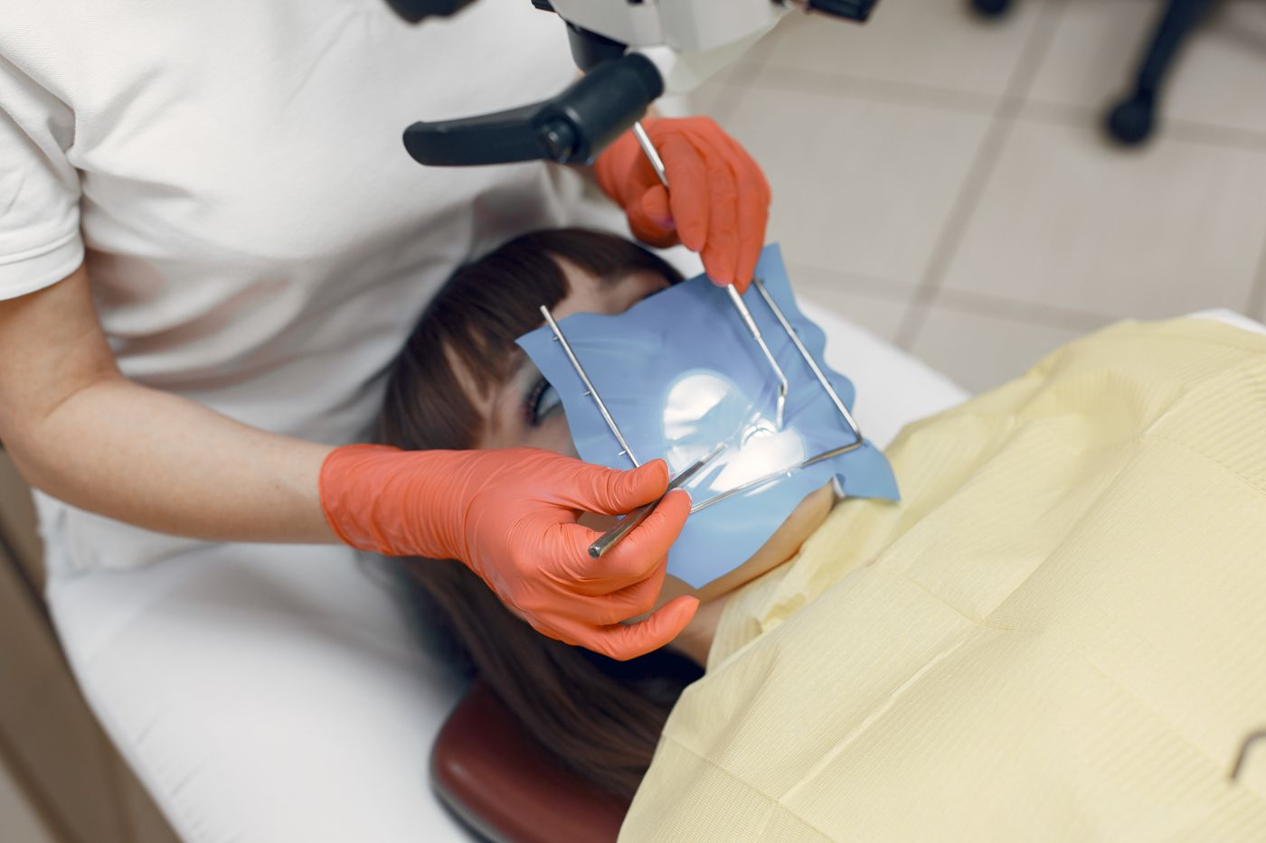
    Magnification is the single biggest change in this procedure over the last twenty years. Working under a microscope allows a much smaller opening, a cleaner cut and a far better seal — see the equipment we use.

    Microsurgical technique is what lifted success rates above 90%
    Microsurgical technique is what lifted reported success rates above 90%.

    Recovery, honestly

    Expect soreness and swelling. It peaks at about 48 hours and then improves daily, and a cold compress in the first day genuinely helps.

    Swelling peaks around 48 hours and settles within a few days
    Swelling peaks around 48 hours, then settles.
    Apicoectomy recovery timeline
    WhenWhat to expectWhat to do
    Day of surgeryNumbness, then soreness as it wears offCold compress, anti-inflammatories, rest
    Day 1–2Swelling peaks; possible bruisingSoft food, no vigorous rinsing or brushing at the site
    Day 3–7Steady improvement; sutures removed around day 7Return to normal activity; gentle cleaning
    Week 2–4Soft tissue fully healedNormal brushing and diet
    Month 3–6Bone fills in at the root tipFollow-up radiograph to confirm healing

    Swipe the table sideways to see every column.

    Soft food and gentle rinsing for the first few days
    Soft food and gentle cleaning for the first few days; most people are back to normal activity the next day.

    Most patients return to work the following day. What takes longer is invisible: the bone defect left by the infection fills in over three to six months, which is why we review with a radiograph rather than judging by symptoms.

    What the success rate really means

    You will find figures ranging from 60% to 97% for this procedure, and the spread is not noise — it is chronology.

    Older studies describe surgery done without magnification, with materials long superseded. Contemporary microsurgical series report around 90% or better, with roughly 97% still successful at five years and more than 75% at ten to thirteen years. If you are reading an alarming number, check when the study was published.

    What it costs

    An apicoectomy typically runs somewhere between a molar root canal and an extraction with an implant. It is billed as a surgical endodontic procedure, which most plans cover at the same percentage as other endodontic work — though it draws on the same annual maximum.

    Ask for a written estimate with your benefits checked before treatment. Our cost guide explains how deductibles and annual maximums interact, and why timing across two plan years sometimes helps.

    The risks worth knowing about

    It is minor surgery, and minor surgery is not no surgery. Bruising and swelling are expected rather than complications.

    The specific risks depend on where the tooth sits. Upper back teeth are close to the maxillary sinus, and lower back teeth to the nerve supplying the lip and chin, so we assess both on 3D imaging before operating rather than discovering the relationship during the procedure. Numbness of the lip is uncommon and usually temporary where it occurs.

    The other honest caveat is that not every case is suitable. A tooth with a vertical root fracture, or too little bone support remaining, will not be improved by surgery — and we would tell you that rather than attempt it.

    Weighing it against extraction

    The honest comparison is not surgery versus doing nothing, but surgery versus removing the tooth and replacing it.

    Bone at the root tip rebuilds over three to six months
    The tooth keeps its own root and ligament, which nothing replaces.

    An apicoectomy keeps your own root, ligament and bone, and costs considerably less than extraction plus an implant. It is also reversible in the sense that extraction remains available afterwards — the reverse is not true. The alternatives compared on ten-year cost sets out the numbers.

    Our apicoectomy procedure page covers the clinical detail, and endodontic surgery the wider category. The published analysis of outcomes after non-surgical treatment fails sets surgery in context against the alternatives.

    Frequently asked questions

    Is an apicoectomy painful?

    During, no more than a root canal — the area is fully anaesthetised. Afterwards expect soreness and swelling for a few days, managed with anti-inflammatories and a cold compress. Most people describe it as comparable to having a tooth out, without losing the tooth.

    How long does it take?

    Between 30 and 90 minutes, depending on which tooth and how accessible the root tip is. Front teeth are usually at the shorter end; lower molars at the longer end.

    What is the success rate?

    Reported success with modern microsurgical technique is around 90% or better, with roughly 97% still doing well at five years in some series. Older figures quoting 60–70% predate the microscope and current materials.

    Can it be repeated if it fails?

    Sometimes, but a second surgery on the same root has a lower success rate. If an apicoectomy fails, extraction and replacement is usually the more sensible next step than a repeat attempt.

    Have Questions or Ready to Schedule?

    Our team is here to help — call, email, or request an appointment online.
    About this guide. Written for patients of Southeast Endodontics, PC, Charleston, SC. Success figures are published averages; whether surgery suits your tooth depends on its anatomy, restoration and treatment history.

  • Root Canals in Children: Baby Teeth and Immature Roots

    Root Canals in Children: Baby Teeth and Immature Roots

    Endodontic treatment in children is not simply the adult procedure scaled down. Their teeth are still developing, and the treatment has to work with that rather than against it.

    Here is what changes with a child’s tooth, why we often treat baby teeth people expect us to pull, and how an unfinished root is handled.

    Key takeaways

    • Baby teeth hold space for the adult teeth behind them — losing one early causes crowding.
    • A pulpotomy treats only the pulp in the crown and leaves the roots alone.
    • An immature permanent tooth has an open root tip that cannot be sealed conventionally.
    • A knocked-out baby tooth should never be replanted.

    Why baby teeth are worth treating

    The instinct to pull a decayed baby tooth is understandable and usually wrong. Primary molars stay in place until around age eleven or twelve, and they are holding the space the permanent premolar will need.

    Remove one early and the adjacent teeth drift into the gap. The permanent tooth then erupts crowded, rotated, or not at all — converting a treatable tooth into years of orthodontics.

    Pulpotomy: the usual treatment for a baby tooth

    Where decay has reached the pulp of a primary tooth but the roots are still healthy, a pulpotomy removes the inflamed tissue in the crown, places a medicated dressing over what remains, and seals it — usually with a stainless steel crown.

    A pulpotomy treats the crown portion of the pulp and leaves the rest
    Only the pulp inside the crown is removed; the root portion stays.

    It is quicker and less invasive than full root canal treatment, and it leaves the root portion of the pulp intact so the tooth resorbs and exfoliates naturally when the time comes.

    Treatment options for children’s teeth, by situation
    SituationUsual treatmentWhat it preserves
    Deep decay, pulp not yet exposedIndirect pulp capThe whole pulp
    Small pulp exposure, healthy pulpDirect pulp cap or partial pulpotomyMost of the pulp
    Inflamed crown pulp, healthy rootsPulpotomy plus crownRoot pulp; natural exfoliation
    Whole pulp necrotic, primary toothPulpectomy with resorbable fillingThe tooth and its space
    Tooth unrestorableExtraction plus space maintainerThe space, not the tooth

    Swipe the table sideways to see every column.

    When the root has not finished forming

    Permanent teeth erupt before their roots are complete. It takes two to three more years for the tip to close, and during that window a tooth is genuinely awkward to treat.

    An immature root has an open tip that has not finished forming
    An immature root has a wide open tip and thin walls.

    The problem is mechanical: there is no narrowing at the root tip to fill against, so conventional sealing is not possible. Thin root walls also make the tooth prone to fracture.

    Approaches to an immature permanent tooth
    ApproachWhen it is usedWhat it achieves
    ApexogenesisPulp still vitalRoot keeps developing normally
    PulpotomyInflamed but vital pulpAround 86% success in immature teeth
    Regenerative endodonticsNecrotic pulp, open apexBest reported root maturation of the three
    ApexificationNecrotic pulp, regeneration unsuitableBuilds a barrier so the canal can be sealed

    Swipe the table sideways to see every column.

    Apexification builds a barrier so the canal can be sealed
    Apexification creates a hard barrier at the tip so the canal can finally be sealed.

    Where the pulp is still alive, keeping it alive is always the goal — a vital pulp continues to lay down dentine and thicken the root walls, which no artificial material replicates.

    Why children’s teeth behave differently

    Two features of a young tooth change the clinical picture entirely, and both work in the child’s favour.

    The pulp chamber is proportionally much larger, which means decay reaches it sooner but also that the pulp has a far richer blood supply and a genuine capacity to heal. Treatments that would be optimistic in an adult are realistic in a child.

    The roots are also still forming, or in the case of baby teeth already resorbing on a schedule. Treatment has to account for where that tooth is in its life cycle rather than treating it as a small adult tooth.

    Trauma is the commonest reason we see children

    Sport, playgrounds and bicycles account for most childhood dental injuries, and upper front teeth take the brunt. Those teeth are often the ones with immature roots, which is why children’s trauma frequently lands with an endodontist.

    Trauma to a developing front tooth is the commonest reason we see children
    Upper front teeth take the impact in most childhood accidents.

    If a permanent tooth is knocked out, the first thirty minutes matter enormously — our avulsed tooth guide covers the protocol. For displaced or chipped teeth, see what to do after an impact. Baby teeth are the exception: never replant one.

    A mouthguard prevents most of these injuries, and our tips for protecting natural teeth cover choosing one.

    What to watch for at home

    Children under-report dental pain, and they adapt to it. A child who has quietly stopped using one side to chew is telling you something even if they say nothing hurts.

    Worth an appointment: a tooth that has changed colour after a knock, a persistent pimple on the gum, swelling of the face or gum, waking at night, or avoiding cold food. Any of those in a child means the same as in an adult.

    After any facial impact, keep watching for months rather than days. A front tooth that darkens six months after a playground fall is the commonest late presentation we see, and the accident is usually long forgotten by then.

    What the appointment is like

    A parent or guardian accompanies every patient under 18, and stays through the consultation. We explain what we are doing in terms the child can follow, because surprise is what frightens children rather than the procedure itself.

    A parent or guardian stays with any patient under 18
    A parent or guardian stays with any patient under 18 throughout.

    Where anxiety is significant, nitrous oxide works well for children and wears off within minutes. Our guide to managing dental anxiety covers the wider options. The pulp-preserving approach used in children is now used selectively in adults too — see vital pulp therapy in mature teeth.

    Keeping a baby tooth until it is ready to go protects the adult tooth behind it
    Keeping a baby tooth until it is ready to go protects the adult tooth behind it.

    If your child has toothache, a discoloured front tooth after a knock, or an injury you are unsure about, it is worth an assessment — contact the practice. The American Academy of Pediatric Dentistry publishes its clinical guideline on pulp therapy for primary and immature permanent teeth.

    Frequently asked questions

    Why treat a baby tooth that is going to fall out anyway?

    Because it is holding space. A primary molar lost early lets the teeth behind it drift forward, and the permanent tooth underneath then has nowhere to erupt. That is an orthodontic problem created to avoid a filling.

    Is a pulpotomy the same as a root canal?

    No. A pulpotomy removes only the inflamed pulp in the crown and leaves the healthy pulp in the roots. A root canal removes all of it. On children’s teeth the partial approach is usually enough and far less invasive.

    What happens if a child knocks out a baby tooth?

    Do not put it back. Replanting a primary tooth can damage the permanent tooth developing above it. See a dentist to check nothing else is injured, but leave the tooth out.

    Can a child have a root canal on a permanent tooth?

    Yes, but if the root is not fully formed it needs a different approach. An open root tip cannot be sealed conventionally, so treatment aims to let the root finish developing or to build a barrier at the tip.

    Have Questions or Ready to Schedule?

    Our team is here to help — call, email, or request an appointment online.
    About this guide. Written for parents by Southeast Endodontics, PC, Charleston, SC. Which treatment suits a child’s tooth depends on their age, the stage of root development and the state of the pulp.

  • Endodontist or General Dentist: Who Should Treat Your Tooth?

    Endodontist or General Dentist: Who Should Treat Your Tooth?

    Your dentist says you need a root canal and offers to do it. Should you say yes, or ask to see a specialist? It is a fair question, and the honest answer depends almost entirely on which tooth it is.

    Here is what actually differs between the two, what the outcome data shows, and the cases where it genuinely matters.

    Key takeaways

    • An endodontist is a dentist plus two to three years of residency limited to this procedure.
    • Reported success is roughly 90–95% for specialists against 85–90% for general practice — though specialists take the harder cases.
    • For a straightforward front tooth, your own dentist is very likely the right choice.
    • Molars, retreatments, unclear diagnoses and “we could not get you numb” are specialist territory.

    What the training difference actually is

    Both are dentists. The difference is what happened afterwards: a residency limited entirely to endodontics, covering diagnosis of orofacial pain, complex canal anatomy, retreatment and surgical endodontics.

    Two to three years of additional training beyond dental school
    Two to three years of full-time residency, after dental school, doing nothing else.

    Volume is the part patients underestimate. A dentist may graduate having completed a handful of root canals; an endodontist typically finishes residency having done several hundred, and then does little else for the rest of their career.

    What the outcome data shows

    Reported root canal outcomes, specialist vs. general practice
    MeasureEndodontistGeneral dentistCaveat
    Reported success rate~90–95%~85–90%Specialists treat harder cases, which understates the gap
    Cases completed in trainingSeveral hundredOften fewer than tenVolume compounds over a career
    Microscope used routinelyStandardLess commonMissed canals are a leading cause of failure
    3D imaging on siteUsualVariesMatters most for molars and retreatment
    Typical fee difference10–20% higherBaselineOften reimbursed at the same rate

    Swipe the table sideways to see every column.

    Read that table carefully in one respect: the success gap is measured across different case mixes. Specialists receive the curved, calcified and previously failed teeth, which drags their average down relative to a like-for-like comparison.

    When your own dentist is the right answer

    Plenty of root canals are genuinely routine, and there is no advantage in paying more for one. Straightforward cases share a profile: a front tooth or premolar, a single canal visible along its whole length on the radiograph, a clear diagnosis, and no history of difficulty getting numb.

    A referral is a judgement about the case, not about your dentist
    A referral is a judgement about the case, not a criticism of your dentist.

    Many general dentists do this work very well and enjoy it. If yours is confident about a case that fits that description, that confidence is usually well founded.

    When it is worth seeing a specialist

    Situations where a specialist referral is worth requesting
    SituationWhy it matters
    Molar, especially a lower first molarThree to four curved canals; the extra canal is easy to miss
    Previous root canal on the toothRetreatment needs the old filling removed before anything else
    You could not get numb last timeHot pulps need supplementary anaesthetic technique
    Nobody is sure which tooth it isReferred pain; treating the wrong tooth is the costly error
    A crack is suspectedDiagnosis relies on magnification and testing, not imaging
    The tooth is calcified on the radiographCanals may be difficult or impossible to locate without a microscope

    Swipe the table sideways to see every column.

    Molars, retreatments and unclear diagnoses are where the difference shows
    Molars, retreatments and unclear diagnoses are where the difference shows up.

    The equipment argument is not marketing. Our page on the technology used here covers it, and why root canals fail explains how often the cause turns out to be anatomy nobody could see.

    Magnification and 3D imaging are standard equipment in a specialist practice
    A missed canal is a leading cause of failure, and magnification is how they are found.

    What a specialist practice does differently

    The equipment matters less than how routinely it is used. A microscope that comes out for difficult cases is not the same as one used on every tooth from the first minute.

    Scheduling differs too. A practice doing only endodontics books the time a molar actually needs rather than fitting it between check-ups, which is why specialist appointments are usually longer and less often split across visits.

    Anaesthesia is the third difference, and the one patients notice. Supplementary techniques for teeth that will not go numb — intraosseous and intraligamentary injections among them — are routine in a specialist practice and less commonly needed in general practice.

    What it costs, honestly

    Expect to pay 10–20% more, commonly $200–$400 extra on a molar. That is a genuine cost and worth weighing.

    Most plans reimburse specialist and general fees at the same rate
    The fee difference is real; the out-of-pocket difference is often smaller.

    Two things reduce it. Most insurance plans reimburse endodontic treatment at the same percentage regardless of who provides it, and a case that has to be redone costs far more than the original difference — the full cost breakdown covers both.

    What happens to the tooth afterwards

    One practical point is worth knowing before you choose, because it surprises people who expect a specialist to handle everything.

    We complete the root canal and place a temporary filling. Your own dentist then places the permanent restoration, which on a back tooth means a crown. So seeing a specialist does not replace your dentist — it adds one appointment in the middle of a course of treatment that returns to them.

    That split is also why the referral note back matters. You should expect a written report and post-treatment radiographs to reach your dentist, so the restorative work can start without repeating anything.

    How to raise it with your dentist

    Ask directly and without awkwardness: how many canals does this tooth have, how confident are you about this one, and would you refer it if it were your own tooth? Those are ordinary clinical questions. So is asking someone else — how a second opinion works sets out where to take which question.

    The gap in reported success is real but smaller than marketing suggests
    The right question is not who is better, but which case this is.

    Dentists refer routinely, and the good ones welcome being asked. If you would rather come to us first, you can — see what a first visit involves and meet Dr. Long and Dr. O’Neal. For colleagues, our guide to referral criteria covers the same ground clinically.

    The American Association of Endodontists sets out its own account of why and when it is worth seeing an endodontist, which is worth reading alongside this page rather than instead of it.

    Frequently asked questions

    Is an endodontist a real dentist?

    Yes. An endodontist completes dental school first, then two to three further years of specialty residency limited to endodontics. It is the same qualification plus a specialty on top, not a different profession.

    Does it cost more to see a specialist?

    Usually 10–20% more per procedure — often $200–$400 on a molar. Most plans reimburse specialist and general fees at the same rate, so the out-of-pocket difference is frequently smaller than the fee difference suggests.

    Do I need a referral?

    Not with us. Many patients arrive by referral from their general dentist, but you can book directly. Either way we send a treatment report back to your dentist so the restorative work can follow.

    Will my dentist be offended if I ask to be referred?

    No, and it is a reasonable question to ask. Referring is a normal part of practice, and most dentists refer the cases they judge to be outside their comfortable range as a matter of routine.

    Have Questions or Ready to Schedule?

    Our team is here to help — call, email, or request an appointment online.
    About this guide. Written by Southeast Endodontics, PC, Charleston, SC. We are a specialist practice, so read the above with that in mind — which is exactly why the section on when your own dentist is the right choice is included.

  • Do You Need a Crown After a Root Canal?

    Do You Need a Crown After a Root Canal?

    The root canal is done, the pain has gone, and now someone is asking you to spend as much again on a crown. It is a fair moment to ask whether it is really necessary.

    For a back tooth, the evidence on this is unusually clear-cut. Here is what the numbers show, and the cases where a crown genuinely is not needed.

    Key takeaways

    • Crowned treated teeth survive at roughly 94% at five years, against 77% without.
    • At ten years the gap widens: about 89% with a crown, 62% without.
    • In one large study, 85% of extracted treated teeth had never received cuspal coverage.
    • Front teeth are the genuine exception — many do not need full coverage.
    • Timing matters: aim for the definitive restoration within about 60 days.

    Why a treated tooth needs protecting

    Two separate things weaken the tooth, and they compound. The decay, fracture or failing restoration that created the problem in the first place has already removed structure, and the access opening needed to reach the canals removes a little more from the biting surface.

    What is lost is not just bulk but bracing. An intact molar resists chewing forces because its walls are connected across the top; once that roof is opened, those walls flex independently every time you bite.

    What the survival data shows

    Survival of root-canal-treated teeth, with and without a crown
    TimeframeWith cuspal coverageWithoutDifference
    5 years~94%~77%17 points
    10 years~89%~62%27 points
    8-year large-cohort survival97% overall85% of extractions had no coverageCoverage dominates the failure data

    Swipe the table sideways to see every column.

    Without cuspal coverage a treated molar splits under normal chewing load
    An uncrowned molar tends to fail by splitting — and a split root cannot be treated.

    The failure mode is what makes this decisive. Uncrowned treated molars typically fail by vertical fracture, and a vertical root fracture is not retreatable — see which cracks can be saved. The tooth is lost, not merely re-treated.

    When a crown is not needed

    This is where blanket advice goes wrong. Front teeth take shearing rather than crushing forces, have a single canal and a much smaller access opening.

    A front tooth with little damage often does not need full coverage
    An incisor with an intact structure and a small access cavity is a different case entirely.
    Which restoration each tooth usually needs
    ToothUsual restorationCrown needed?
    Incisor, intact walls, small accessBonded composite restorationOften not
    Incisor with large existing restoration or discolourationCrown or veneerUsually
    PremolarCuspal coverage restorationUsually
    MolarFull-coverage crown or onlayAlmost always
    Any tooth with a cracked cuspFull coverageYes

    Swipe the table sideways to see every column.

    Timing, and why it matters

    A temporary filling is exactly that. It seals well for a few weeks and then begins to leak, and leakage lets saliva and bacteria back into canals that were just disinfected.

    Outcomes are better when the crown follows within about 60 days
    Outcomes improve when the definitive restoration follows within about two months.

    That is one of the routes to treatment failing and needing retreatment — not because the root canal was poor, but because the seal above it was lost. Book the restorative appointment before you leave, not once the tooth starts bothering you again.

    Crown, onlay, or filling?

    A crown is not the only way to get cuspal coverage. An onlay covers the cusps while preserving more natural tooth, which is often the better choice where the walls are still sound.

    A filling restores the hole; a crown redistributes the load
    A filling restores the shape; cuspal coverage changes how force travels through the tooth.

    The distinction that matters is not crown versus onlay but covered versus not covered. A large direct filling in a treated molar leaves the walls unbraced, which is precisely the situation the survival data warns about.

    What actually happens if you delay

    Two separate failures compete for the tooth, and they run on different clocks.

    The slower one is leakage. A temporary filling wears and begins to admit saliva within weeks, recontaminating the canal system underneath. This is silent — you will not feel it happening, and it surfaces months later as a tooth that needs retreating.

    The faster one is fracture. Every bite flexes unbraced walls, and it takes one awkward bite on something hard to split a cusp. If that split runs below the gumline the tooth is lost outright, which is what the survival figures above are really describing.

    Do you need a post as well?

    Posts are widely misunderstood as strengthening a tooth. They do not — a post exists to retain a core build-up where too little coronal structure remains to hold one on its own.

    Molars usually manage without, because the pulp chamber itself retains the core. Where a post is genuinely needed it should be as conservative as possible, since preparing the canal to receive one removes dentine and raises the risk of root fracture.

    Who does it, and what it costs

    We complete the root canal and place a temporary filling; your own general dentist then places the definitive restoration. That split in responsibility surprises a lot of patients, and it is worth knowing about before you set a budget.

    The crown is usually made by your general dentist, not by us
    The crown is usually made by your general dentist’s laboratory, not by us.

    Expect $800–$2,000 for the crown on top of the treatment fee — the full cost breakdown covers how insurance annual maximums affect the total, and why splitting the two across plan years can help.

    If the total is genuinely out of reach, say so before treatment rather than after. A root canal on a molar that never receives cuspal coverage is, on the survival data, an expensive route to the same extraction you were trying to avoid.

    Crowned treated teeth survive at roughly 94% at five years, against 77% without
    Treated and properly restored, the tooth is simply a tooth again.

    That is not an argument for extracting teeth cheaply. It is an argument for having the whole cost on the table at the start, so the decision you make is the one you would still make a year later. Restoration quality also predicts survival as strongly as the root filling does — see what affects the outcome.

    Restored properly, a treated tooth needs nothing special — brush, floss and keep your check-ups. What is normal in the first week covers the period before the crown goes on. The published eight-year survival analysis of treated teeth by restoration type is the source for the figures above.

    Frequently asked questions

    Do all root-canal-treated teeth need a crown?

    No. Back teeth almost always do, because they take heavy chewing load and have lost internal structure. A front tooth with a small access opening and otherwise intact enamel is often fine with a bonded filling.

    How long can I leave it before getting the crown?

    Weeks, not months. Outcomes are better when the definitive restoration follows within about 60 days. A temporary filling is not designed to seal long term, and leakage lets bacteria back into canals that were just cleaned.

    Will I need a post as well?

    Often not. Molars usually retain a core restoration in the pulp chamber without one. Posts are mainly for teeth with very little coronal structure left, and they carry their own risk of root fracture.

    What if I cannot afford the crown right now?

    Say so before treatment starts. If the budget genuinely covers only one thing, that changes the conversation about whether the tooth is the right investment — a root canal without a crown on a molar frequently ends in extraction anyway.

    Have Questions or Ready to Schedule?

    Our team is here to help — call, email, or request an appointment online.

    About this guide. Written for patients of Southeast Endodontics, PC, Charleston, SC. Survival figures are from published studies of large cohorts; your dentist decides the right restoration for your particular tooth.

  • Are Root Canals Safe? What the Evidence Actually Says

    Are Root Canals Safe? What the Evidence Actually Says

    Search “are root canals safe” and you will find pages claiming they cause cancer, heart disease and chronic fatigue. Those claims share a single origin, and it is worth knowing what it is.

    This is where the idea came from, why it did not survive scrutiny, and what the evidence actually supports — including the risks that are real.

    Key takeaways

    • The “root canals cause disease” claim traces to one researcher’s work in the 1920s.
    • Those experiments had no controls and have never been reproduced.
    • No controlled study since has found a link to systemic disease.
    • Extracting a restorable tooth is not the safer option — it carries its own risks.

    Where the claim comes from

    In the 1910s and 1920s, “focal infection theory” held that a hidden infection anywhere in the body could cause disease elsewhere in it. The idea was taken seriously at the time and used to justify removing tonsils, and healthy teeth, on a very large scale across Europe and the United States.

    Weston Price's experiments lacked controls and could not be reproduced
    The focal infection theory dates from an era before antibiotics and before modern immunology.

    Weston Price extracted root-canal-treated teeth from chronically ill patients and implanted them under the skin of rabbits. When the rabbits sickened, he concluded the teeth had caused the patients’ illnesses.

    Why that research does not hold up

    Judged by any modern standard, the work fails on basics:

    • No control group. Healthy extracted teeth were not implanted for comparison.
    • No sterile technique. Teeth were contaminated during extraction and handling, so whatever the rabbits reacted to may never have been in the tooth.
    • Enormous relative dose. A whole human tooth under a rabbit’s skin is not comparable to a treated tooth in a human jaw.
    • Correlation read as causation. Chronically ill people in that era had more dental disease for reasons unrelated to root canals.
    • Never reproduced. A century of attempts has not replicated the findings.
    The claim, and what the evidence shows
    ClaimOriginWhat research since has found
    Root canals cause cancerFocal infection theory, 1920sNo association found in controlled studies
    Treated teeth harbour dangerous bacteriaPrice’s rabbit experimentsBacteria exist in many body sites without causing systemic disease
    Extraction is the safer optionFollows from the aboveExtraction has its own surgical risks and consequences
    Dentists suppress thisDocumentary claims, 2019The theory was abandoned because it failed testing, publicly

    Swipe the table sideways to see every column.

    What the modern evidence says

    Focal infection theory was not suppressed — it was investigated and abandoned as better methods emerged. Antibiotics, controlled trials and modern immunology all arrived after Price’s work and none supported it.

    Decades of controlled research have found no link to systemic disease
    The theory was not buried. It was tested, repeatedly, and did not hold.

    The professional consensus is unambiguous: there is no valid evidence linking endodontically treated teeth to systemic disease. Our page on root canal safety and the common myths cover the same ground from the clinical side.

    The risks that are real

    Being straight about this matters more than reassurance. Genuine risks exist, and they are ordinary clinical ones:

    • Treatment can fail. Around 86% of initial treatments succeed, so roughly one in seven needs something further — see retreatment and surgery.
    • The tooth can fracture if it is not properly restored, which is why a crown matters on back teeth.
    • Post-operative discomfort for a few days is normal and expected.
    • Rarely, an instrument separates in a canal, or an existing crack is discovered mid-treatment that changes the prognosis.

    How to weigh a health claim you find online

    This topic is a useful test case, because the same pattern recurs across health misinformation generally.

    • Check the date of the underlying research, not the date of the article quoting it. A 2019 documentary citing 1920s experiments is still 1920s evidence.
    • Ask whether it was reproduced. A single striking result nobody has replicated in a century is a red flag, not a suppressed truth.
    • Look for the control group. Without one, a study cannot separate the effect being claimed from everything else going on.
    • Notice what is being sold. Claims that root canals are toxic frequently arrive alongside an offer to extract teeth and replace them.

    None of that requires a clinical background. It is the same reasoning you would apply to any other claim about your health.

    What about the X-rays?

    Endodontic diagnosis needs imaging, and people reasonably ask about the dose. It is small — a fraction of the background radiation you absorb from the environment in a normal year.

    A full endodontic series delivers a small fraction of your annual background dose
    Digital sensors cut the dose substantially against the film they replaced.

    The alternative is worse. Treating a molar without seeing its root anatomy is how canals get missed, and a missed canal is a far more likely source of harm than the exposure.

    Treating versus extracting, on risk

    Risks of root canal treatment compared with extraction
    ConsiderationRoot canal treatmentExtraction
    Procedure typeNon-surgical in almost all casesSurgical, with bleeding and healing
    Bone preservedYes — the root maintains itNo — resorption begins immediately
    Adjacent teeth affectedNoDrift and over-eruption over years
    Further treatment likelyCrown; occasionally retreatmentImplant or bridge to fill the gap
    If it failsRetreatment or surgery availableNothing to retreat — the tooth is gone

    Swipe the table sideways to see every column.

    The comparison that actually matters

    “Is a root canal safe?” is the wrong question on its own. The real question is whether it is safer than the alternative for your tooth, and for a restorable tooth the answer is consistently yes.

    Extracting a healthy-rooted tooth carries more risk than treating it
    Nothing replaces a natural tooth root, its ligament and the bone it maintains.

    Extraction is surgery. It leaves a gap that drives bone loss and tooth movement unless replaced, and replacement means an implant or bridge with their own risks — compared here on ten-year cost and outcome.

    Isolation and sterile technique are what make the procedure predictable
    Isolation, sterile instruments and magnification are what make outcomes predictable.

    If something you have read worries you, raise it at your consultation — see what a first visit involves. The American Association of Endodontists publishes its position on focal infection theory with the underlying references.

    Bring the claims you have read — they are worth talking through properly
    Bring what you have read. It is a reasonable conversation to want to have.

    Frequently asked questions

    Do root canals cause cancer or chronic disease?

    No. That claim comes from research done in the 1920s using methods that would not pass review today, and it has not been reproduced since. Decades of controlled studies have found no link between endodontically treated teeth and systemic disease.

    Where did the “root canals are toxic” idea come from?

    From Weston Price, who implanted extracted teeth under the skin of rabbits and concluded that the teeth caused the illnesses that followed. The experiments had no controls, no sterile technique, and predate modern microbiology by decades.

    Is it safer to just extract the tooth?

    No. Extraction is a surgical procedure with its own risks, and it leaves a gap that has to be managed. Nothing about removing a restorable tooth is inherently safer than treating it.

    How much radiation is involved?

    Very little. The imaging used for endodontic diagnosis delivers a small fraction of the background radiation you receive from the environment each year, and we use it because treating without seeing the anatomy is the greater risk.

    Have Questions or Ready to Schedule?

    Our team is here to help — call, email, or request an appointment online.
    About this guide. Written for patients of Southeast Endodontics, PC, Charleston, SC. It summarises the published position of the endodontic specialty; it is not a substitute for discussing your own health circumstances with your clinicians.

  • What Actually Happens During a Root Canal

    What Actually Happens During a Root Canal

    Most of what people dread about a root canal comes from not knowing what happens during one. The reality is closer to a long filling than to surgery.

    Here is the whole appointment, step by step, with honest answers on how each part feels and how long it takes.

    Key takeaways

    • The tooth is fully numb throughout — it feels much like having a filling done.
    • Most appointments run 60–120 minutes, driven by how many canals the tooth has.
    • The rubber dam is there to keep saliva out, which is what makes the result last.
    • You leave with a temporary filling; the permanent restoration comes later.

    Before anything starts

    Nothing happens on the first visit until we know what is wrong and which tooth is causing it. That sounds obvious, and it is the step most often skipped when treatment is rushed.

    We test the tooth and its neighbours, because pulpal pain refers readily between upper and lower teeth on the same side. Imaging then shows the root anatomy and how much infection has reached the bone.

    Only after that do we confirm what the tooth needs. Sometimes the answer is that it does not need a root canal at all — see what different sensitivity patterns mean.

    Step by step, what actually happens

    1. Numbing (10–15 minutes). Topical gel, then local anaesthetic. We test that the tooth is numb before starting — tell us if it is not.
    2. The dam goes on. A thin sheet isolates the tooth so it stays dry and clean.
    3. Access. A small opening is made through the biting surface to reach the pulp chamber.
    4. Cleaning and shaping. The infected tissue is removed and each canal is cleaned, shaped and disinfected.
    5. Filling and sealing. The canals are dried and filled with gutta-percha, then sealed so bacteria cannot re-enter.
    6. Temporary filling. The access opening is closed until your dentist places the permanent restoration.
    Getting the tooth properly numb is the step everything else depends on
    Topical gel first, then the injection. Getting the tooth properly numb is the step everything depends on.

    Why the rubber dam matters more than it looks

    Patients often ask about the dam because it looks like the most intrusive part. It is actually the step that most affects whether treatment lasts.

    The dam keeps the tooth dry and stops anything reaching the back of your throat
    Isolation is not a formality — saliva reaching a cleaned canal undoes the work.

    Saliva is full of bacteria. Cleaning a canal thoroughly and then letting saliva into it recontaminates everything, which is one route to treatment failing later. The dam also means nothing can be dropped toward your throat.

    The part that takes the time

    Cleaning is where the appointment is spent. A canal is not a smooth tube — it branches, curves, and narrows, and every part of that system has to be disinfected.

    Each canal is cleaned, shaped and disinfected in turn
    Each canal is treated in turn, which is why the number of canals drives the appointment length.
    What drives how long your appointment takes
    ToothCanals, typicallyTypical appointmentWhy
    Front tooth145–60 minutesSingle straight canal, easy access
    Premolar1–260–90 minutesTwo canals in many cases
    Molar3–4+90–120 minutesMultiple curved canals, harder access
    Any tooth with acute infectionVariesMay need two visitsMedication placed between appointments
    RetreatmentVariesLonger than the originalOld filling material must come out first

    Swipe the table sideways to see every column.

    The cleaned canals are sealed with gutta-percha so bacteria cannot return
    Once the canals are clean and dry, they are filled and sealed in the same visit.

    Technology changes this part. Magnification finds canals that would otherwise be missed, and fluid-based cleaning reaches side branches instruments cannot — see how GentleWave compares with conventional treatment and the equipment we use.

    What you will notice, and what you will not

    Sensations during treatment: what is expected and what to report
    What you noticeExpected?What to do
    Pressure and vibrationYesNormal — the tooth is numb to pain, not to pressure
    The sound of instrumentsYesHeadphones help; ask before you start
    Jaw ache from holding openYesSay so — we can pause or use a bite prop
    A sharp twingeNoRaise your hand immediately; more anaesthetic is needed
    Tasting the irrigating solutionNoTell us — the dam seal may need adjusting
    Feeling unable to swallowNoSignal; the dam can be released in seconds

    Swipe the table sideways to see every column.

    How it actually feels

    Numb. That is the honest answer. You will feel pressure and vibration, and hear the instruments, but the tooth itself should feel nothing.

    Most appointments run 60–120 minutes depending on the tooth
    Plan for around two hours at the practice for a molar, including checks and paperwork.

    The exception worth naming: an acutely inflamed pulp is harder to anaesthetise, so a “hot” tooth occasionally needs supplementary technique. Say something the moment you feel anything sharp — that is information we need, not an inconvenience.

    If anxiety rather than pain is the concern, the sedation options are worth reading before you book, and our pre-treatment instructions cover the practical preparation.

    When a second visit is needed

    Most treatment is completed in a single appointment. Two situations change that, and neither means anything has gone wrong.

    The first is significant infection. Where there is swelling or persistent drainage, we place an antimicrobial dressing, seal the tooth temporarily, and let things settle for a week or two before filling the canals. Sealing an actively discharging canal simply traps the problem inside.

    The second is complexity. A molar with four curved canals, or a case where an unexpected extra canal turns up, is sometimes better finished properly at a second visit than rushed at the end of a long first one. We will tell you before you leave which of these applies to you.

    Leaving, and what comes next

    You can normally drive yourself home and go straight back to work if you want to. The numbness wears off gradually over two to four hours; avoid chewing on that side until it has completely gone.

    A temporary filling protects the tooth until the permanent restoration
    You leave with a temporary filling — the permanent restoration is a separate appointment.

    The tooth is not finished yet. A temporary filling is designed for weeks, and a back tooth needs a crown to stop it fracturing. What is normal in the first week covers the days ahead, and Delta Dental’s step-by-step walkthrough of the procedure is a useful second read. In some cases the whole pulp does not need removing at all — see when the nerve can be kept alive instead.

    Frequently asked questions

    Does a root canal hurt?

    The procedure itself feels much like having a filling, because the tooth is fully anaesthetised. Nearly all the pain associated with root canals is the pain that brings people in — treatment is what ends it.

    How long does a root canal take?

    Most appointments run 60 to 120 minutes. A front tooth with one canal is at the short end; a lower molar with four curved canals is at the long end. Cases with significant infection sometimes need a second visit.

    Will I be awake?

    Yes. You are numb, not asleep. If the appointment itself is what worries you, sedation options are available and worth asking about in advance rather than on the day.

    Why is a rubber sheet put over the tooth?

    The dam keeps the tooth dry and free of saliva, which matters because bacteria in saliva would recontaminate the canals. It also stops small instruments or irrigating solutions reaching the back of your throat.

    Have Questions or Ready to Schedule?

    Our team is here to help — call, email, or request an appointment online.
    About this guide. Written for patients of Southeast Endodontics, PC, Charleston, SC. Appointment times are typical rather than promised — anatomy varies more than most people expect.

  • Failed Root Canal: Retreatment, Surgery or Extraction?

    Failed Root Canal: Retreatment, Surgery or Extraction?

    A root canal is supposed to end the problem. When the tooth starts aching again — six months later, or six years — it feels like the treatment was wasted.

    Usually it was not. Most previously treated teeth can still be saved, and there is a clear order to the options. Here is why treatment fails, what each second attempt actually achieves, and when stopping is the right call.

    Key takeaways

    • Initial treatment succeeds around 86% of the time, so failures happen without anyone doing anything wrong.
    • The usual cause is anatomy that was never reached — a missed canal or an incomplete seal.
    • Non-surgical retreatment comes first, then surgery, then extraction. In that order.
    • Microsurgical apicoectomy reports success rates of 90% or better, well above older figures.

    Why root canals fail

    Failure almost always comes down to bacteria surviving somewhere in the canal system, or getting back in afterwards.

    Returning pain, a gum bump, or swelling are the usual signs
    Returning pain months or years later is the most common way failure announces itself.
    • A missed canal. Molars often have an extra canal that is genuinely hard to find without magnification.
    • Complex anatomy — curves, branches and fins that instruments cannot reach.
    • An incomplete seal at the root tip, letting bacteria persist. This is the single most cited cause.
    • Delayed restoration. A temporary filling left too long leaks, and bacteria re-enter a cleaned canal.
    • New decay or a fracture creating a fresh route in, years after successful treatment.

    The signs to take seriously

    A treated tooth should be comfortable. Any of these warrants an assessment rather than watchful waiting:

    • Pain returning after a period of comfort, particularly on biting.
    • A pimple or bump on the gum near the tooth, which may drain and taste unpleasant.
    • Swelling or tenderness over the root.
    • The tooth darkening compared with its neighbours.
    • An area of shadow at the root tip that your dentist notices on a routine radiograph, even with no symptoms.

    That last one matters — a failing tooth is often silent. Symptoms overlap with a crack in the tooth, which is why testing rather than guesswork decides it.

    What each sign usually points to
    What you noticeMost likely causeWhat it changes
    Pain on biting that returns after months of comfortMissed canal, or a crack developingNeeds testing before assuming retreatment
    A bump on the gum that drainsPersistent infection at the root tipRetreatment or apicoectomy
    Swelling over the root, no painChronic low-grade infectionUsually still savable
    Shadow at the root tip on a routine X-rayIncomplete healing, often symptomlessAssessment now, before it flares
    Tooth feels looseBone loss or a root fracturePrognosis is poorer; may mean extraction

    Swipe the table sideways to see every column.

    Your three options, compared

    Retreatment vs. apicoectomy vs. extraction: what each involves
    OptionWhat happensReported successBest when
    Non-surgical retreatmentTooth reopened, old filling removed, canals recleaned and resealed~78% overall; 71–77% at 1–5 yearsThe canals can be re-accessed — the usual first choice
    Apicoectomy (root-end surgery)Root tip and infected tissue removed through the gum, root end sealed90%+ when done microsurgicallyA post or crown blocks re-entry, or retreatment has failed
    Extraction + implantTooth removed, implant placed after healing~90% implant survival at ~7 yearsThe tooth is fractured or unrestorable
    Extraction aloneTooth removed, gap leftn/aRarely ideal — see the alternatives guide

    Swipe the table sideways to see every column.

    Weighing retreatment, surgery and extraction with your endodontist
    The right answer depends on why it failed, not just that it failed.

    Retreatment: the usual first step

    Retreatment means undoing the first attempt before redoing it. The crown or filling is opened, the previous root filling material removed, and the canal system explored properly under a microscope.

    Retreatment means reopening the tooth and cleaning what was missed
    Magnification is what makes finding a missed canal realistic rather than lucky.

    Modern imaging changes the odds here. Cone-beam CT shows canals a standard radiograph flattens into invisibility, which is often exactly where the problem was hiding.

    3D imaging often reveals the canal that was missed first time
    3D imaging frequently finds the canal a two-dimensional film could never show.

    Our page on endodontic retreatment covers the procedure, and the technology we use explains the imaging.

    When surgery is the better route

    Sometimes reopening the tooth is the wrong move — a well-fitting post and crown may not survive removal, or the canal may be blocked by a separated instrument.

    An apicoectomy seals the root tip from outside when retreatment cannot reach it
    An apicoectomy works from outside the tooth, sealing the root tip directly.

    An apicoectomy approaches from the side instead: the root tip and surrounding infected tissue are removed through the gum, and the end of the root is sealed. Done under a microscope, reported success is 90% or better. See how apicoectomy works and endodontic surgery generally.

    How long before you know it worked

    Retreatment is not judged on how the tooth feels next week. Symptoms usually settle within days, but the bone around the root tip rebuilds slowly.

    We normally review at six months and again at a year, comparing radiographs to see whether the shadow is shrinking. A tooth that is comfortable but shows no bone healing at twelve months is the one that may still need surgery.

    That timescale is worth knowing before you start, because it changes how you read the first few weeks. Discomfort easing is a good sign; it is not yet proof, and neither is a single follow-up film.

    When to stop trying

    Being honest about limits matters as much as knowing the options. We will recommend extraction when the tooth has a vertical root fracture, when too little structure remains to restore it, when severe bone loss has already loosened it, or when both retreatment and surgery have been tried.

    A second opinion is worth having before agreeing to extraction
    Get the assessment before the irreversible decision, not after.

    The order matters because it runs from least to most irreversible. Once the tooth is out it cannot be put back, so a specialist opinion belongs before that decision. The alternatives compared on ten-year cost covers what follows extraction.

    Most previously treated teeth can still be saved
    Most previously treated teeth can still be kept.

    If you have been told a treated tooth is hopeless, that is worth a second look. Our guide to getting a second opinion on a tooth covers how to get your records and what to ask. Dr. Long and Dr. O’Neal assess these cases routinely — see what a consultation involves. The published 13-year analysis of outcomes after non-surgical treatment fails is the underlying evidence for the ordering above.

    Frequently asked questions

    How common is it for a root canal to fail?

    Uncommon, but not rare. Published figures put initial treatment success around 86%, so roughly one tooth in seven eventually needs something further — sometimes years later.

    Can a failed root canal be fixed without surgery?

    Usually yes. Non-surgical retreatment is the first option: the tooth is reopened, the old filling material removed, missed anatomy cleaned, and the canals resealed. Surgery is generally reserved for cases where that is not possible or has already been tried.

    Does retreatment hurt more than the first time?

    No. It takes longer, because the previous filling has to come out before anything else can happen, but it is done under the same anaesthetic and most patients find it comparable.

    Should I just have an implant instead?

    Not as a first move. Retreatment is less invasive, cheaper, and keeps your own root and ligament. An implant is a reasonable answer once the tooth genuinely cannot be saved — but that decision is irreversible, so it is worth an endodontist’s opinion first.

    Have Questions or Ready to Schedule?

    Our team is here to help — call, email, or request an appointment online.

    About this guide. Written for patients of Southeast Endodontics, PC, Charleston, SC. Success rates are published averages across many studies; your tooth’s prognosis depends on its own anatomy and history.