A standard dental X-ray compresses a three-dimensional root into a flat picture. Most of the time that is enough. Occasionally the thing you most need to see is hidden precisely because of that flattening.
Here is what cone-beam imaging shows that a film cannot, when it is genuinely worth taking, and why it should not be routine.
Key takeaways
- A conventional radiograph superimposes structures; a scan separates them into three planes.
- A limited field-of-view scan costs roughly the dose of two periapical radiographs.
- It is used selectively — the professional position is explicitly against routine use.
- The commonest findings are extra canals, resorption, and the true extent of infection.
What a flat image hides
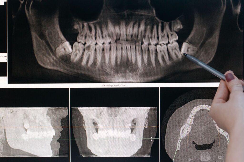
A periapical radiograph is a shadow. Everything between the source and the sensor is projected onto one plane, so structures sitting behind one another overlap.
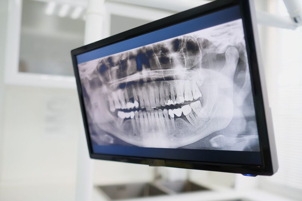
In a molar with four canals, two frequently lie directly behind the others from the angle the image is taken. They are not faint on the film — they are invisible, hidden behind the roots in front. Bone loss also has to reach a certain extent before it registers at all, so early changes at a root tip can be genuinely undetectable.
What a scan adds
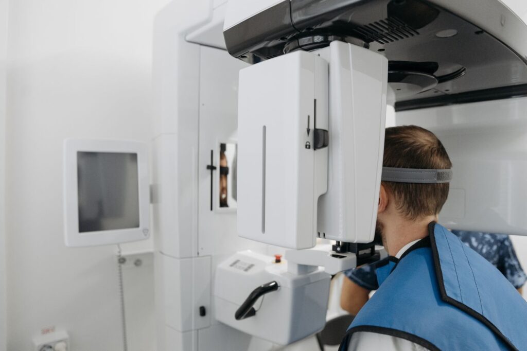
Cone-beam imaging takes a series of images as the machine rotates, then reconstructs them into a volume that can be viewed in cross-section from any angle.
| Question | Standard radiograph | 3D scan |
|---|---|---|
| How many canals does this tooth have? | Often incomplete | Reliable, including canals hidden behind others |
| How far has the infection spread? | Underestimates early lesions | Shows true extent in three dimensions |
| Is there resorption, and how far around? | Frequently missed | Shows position and circumferential spread |
| Where is the sinus or nerve relative to the root? | Approximate | Precise — important before surgery |
| Is this root fractured? | Rarely visible | Sometimes visible; still not guaranteed |
| Routine diagnosis of a straightforward tooth | Entirely adequate | Unnecessary |
Swipe the table sideways to see every column.
That last row matters as much as the others. For most teeth a conventional radiograph answers the question completely, and taking a scan adds dose without adding information.
When it is actually indicated
The joint position of the endodontic and oral radiology specialties is that scans should be used selectively, guided by the clinical question. In practice that means:
- Retreatment, where the reason the first attempt failed is not visible — see why root canals fail.
- Suspected extra canals, particularly in upper molars.
- Suspected resorption, where extent decides whether the tooth is restorable — see how resorption is assessed.
- Before surgery, to establish exactly where the sinus floor or the nerve to the lip sits relative to the root tip.
- Dental trauma, where root fractures and displacement are hard to assess on a film.
- Persistent symptoms with no explanation on conventional imaging.
What we actually find
Scans are worth taking only where the finding changes something. In endodontic practice a handful of findings recur.
| Finding | How often it matters | What changes |
|---|---|---|
| An untreated canal in a previously treated tooth | The commonest useful finding | Retreatment becomes worthwhile instead of extraction |
| A lesion larger than the film suggested | Frequent | Prognosis and follow-up interval |
| Resorption, and how far around the root | Occasional but decisive | Whether the tooth is restorable at all |
| Root tip within or against the sinus | Common in upper molars | Surgical approach, and what we warn you about |
| Nerve canal close to a lower root | Common in lower molars | Whether surgery is advisable |
| Nothing unexpected | Often | Confirms the original plan — still useful |
Swipe the table sideways to see every column.
The last row deserves as much weight as the first. A scan that confirms there is nothing hidden is not a wasted scan; it converts a suspicion into a decision, and it is frequently what allows treatment to proceed with confidence rather than caution.
It also occasionally shows something outside the tooth entirely — sinus disease, or a lesion unrelated to the tooth we were investigating. Where that happens we refer it on rather than treating around it.
The dose question, answered properly
A scan is more radiation than a single film. That is worth stating plainly rather than glossing.
The relevant comparison, though, is not scan versus nothing. It is scan versus the several conventional images at different angles that would otherwise be taken trying to answer the same question — and often failing. A limited field-of-view scan is roughly equivalent to two periapical radiographs, and in a complex case it can represent a net reduction.
The governing principle is ALARA — as low as reasonably achievable. That means the smallest field of view that answers the question, taken only when the answer will change what we do. It also means we do not take one just because the machine is there.
Where it changes the plan
Imaging is only worth taking if it can change a decision. In endodontics it regularly does.
A scan showing an untreated canal converts a tooth destined for extraction into one that can be retreated. A scan showing resorption wrapped around most of a root converts an optimistic repair plan into an honest conversation about the alternatives. Before root-end surgery, it turns an estimate about where the nerve runs into a measurement. It also earns its place in heavily calcified teeth, which is why it comes up so often in treatment planning for older patients.
What it is like to have one
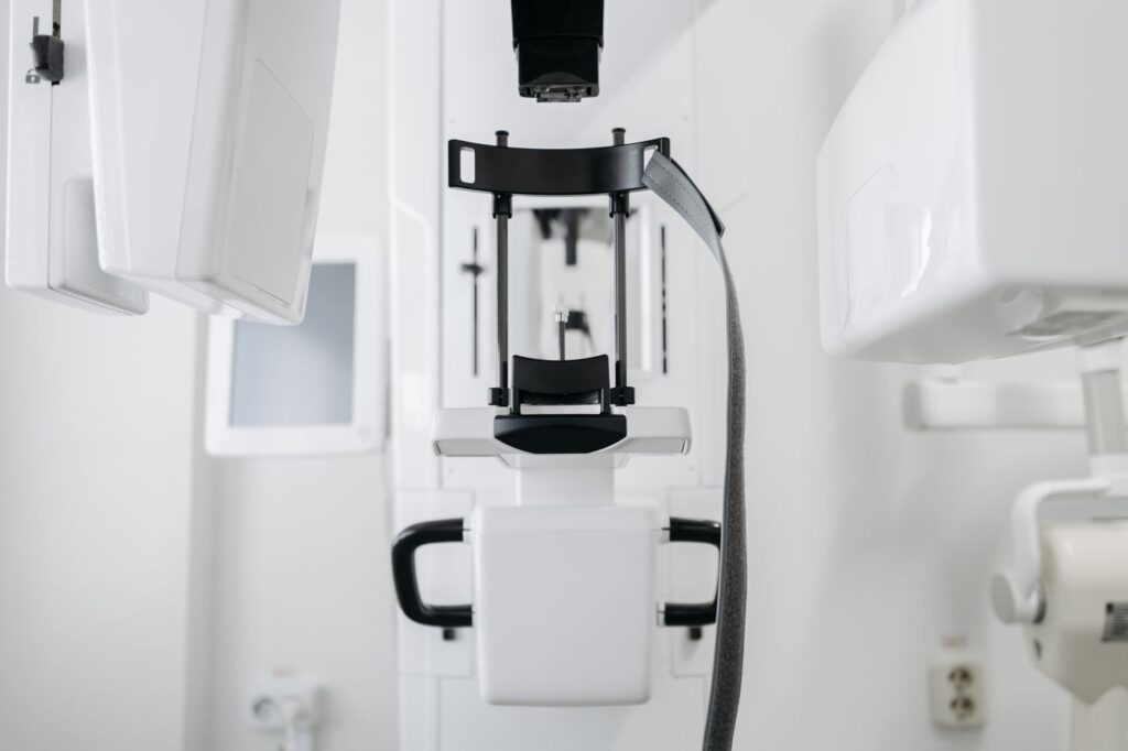
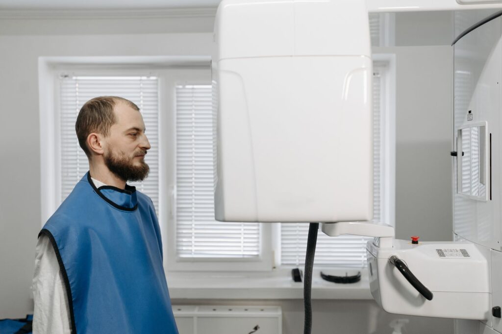
Undramatic, which is rather the point of mentioning it. You sit or stand with your chin resting on a support while the arm rotates once around your head. Ten to twenty seconds, no enclosed tube, nothing injected, no dye to swallow, and you can be spoken to throughout.
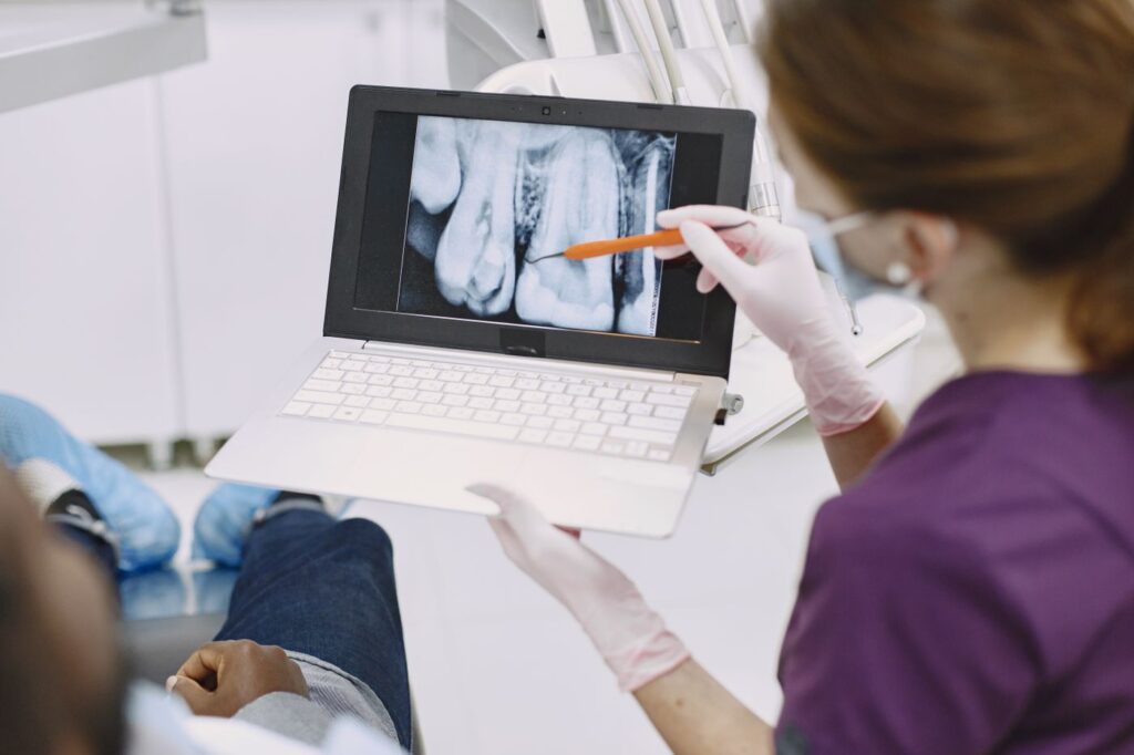
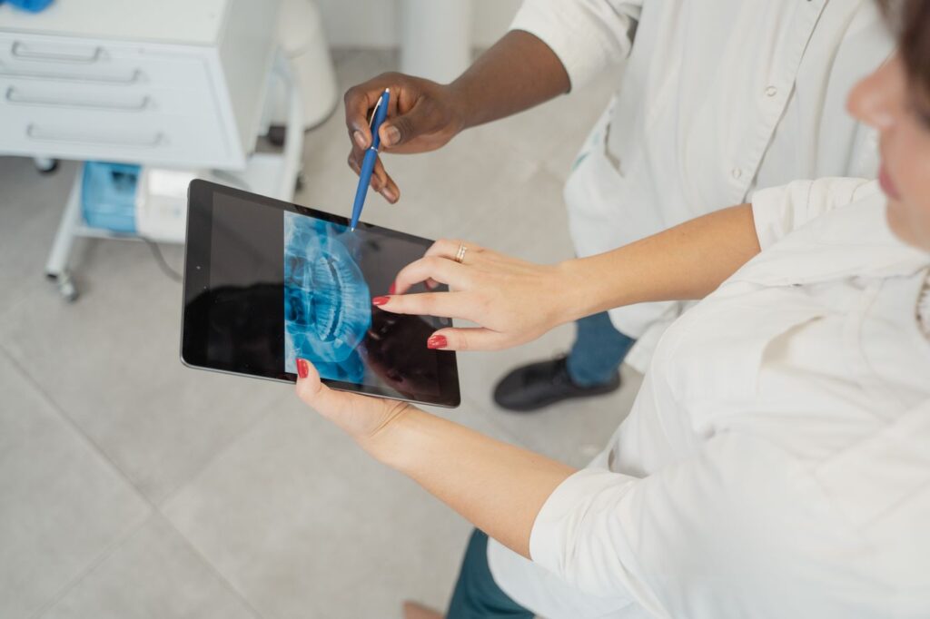
We review the volume with you where it helps — seeing an extra canal on screen explains a treatment plan faster than any description. Our page on the technology used in the practice covers the rest of the equipment, and the joint position statement on cone-beam imaging in endodontics sets out the professional criteria.
Frequently asked questions
Is a 3D scan more radiation than an X-ray?
Yes, but less than people assume. A limited field-of-view endodontic scan delivers roughly the dose of about two standard periapical radiographs, and it can save taking several conventional images in a complex case.
Will I need one?
Probably not. Most root canal treatment is planned from standard radiographs. Scans are taken selectively — for retreatment, suspected extra canals, resorption, trauma, or before surgery.
What does the scan feel like?
Nothing. You sit or stand still while the machine rotates around your head for ten to twenty seconds. There is no enclosed tube and no injection.
Can my dentist see the scan too?
Yes. Where a scan informs the treatment plan, the findings go into the report we send back to your dentist, along with post-treatment radiographs.
Have Questions or Ready to Schedule?
Our team is here to help — call, email, or request an appointment online.