10-E Farmfield Avenue, Charleston, SC 29407

235-213-0595

Category: Dental Technology

The technology behind modern endodontics, and what it changes for patients.

  • What a 3D Scan Shows That an X-Ray Cannot

    What a 3D Scan Shows That an X-Ray Cannot

    A standard dental X-ray compresses a three-dimensional root into a flat picture. Most of the time that is enough. Occasionally the thing you most need to see is hidden precisely because of that flattening.

    Here is what cone-beam imaging shows that a film cannot, when it is genuinely worth taking, and why it should not be routine.

    Key takeaways

    • A conventional radiograph superimposes structures; a scan separates them into three planes.
    • A limited field-of-view scan costs roughly the dose of two periapical radiographs.
    • It is used selectively — the professional position is explicitly against routine use.
    • The commonest findings are extra canals, resorption, and the true extent of infection.

    What a flat image hides

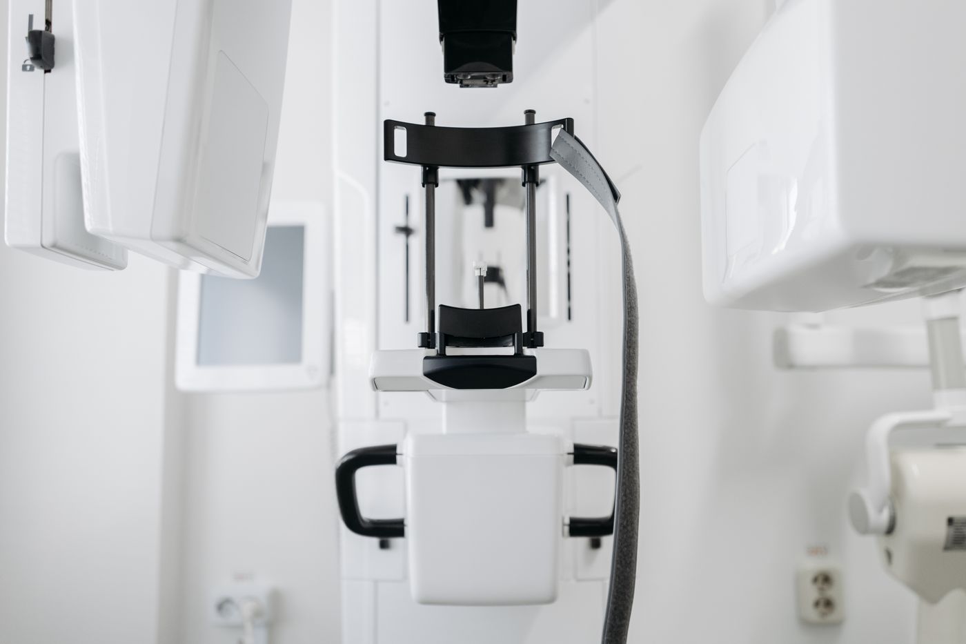
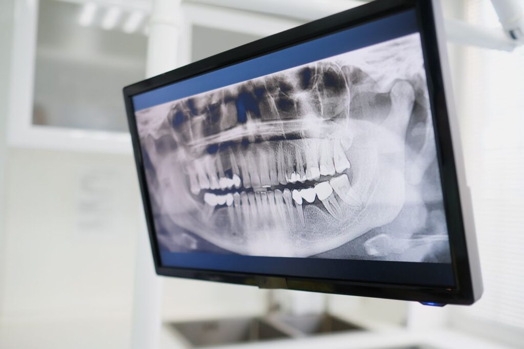
    A periapical radiograph is a shadow. Everything between the source and the sensor is projected onto one plane, so structures sitting behind one another overlap.
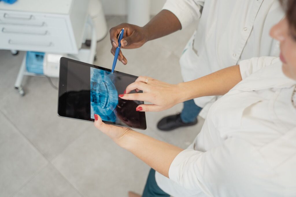
    A standard radiograph compresses a three-dimensional root into a flat image
    A standard radiograph compresses a three-dimensional root into a single plane.

    In a molar with four canals, two frequently lie directly behind the others from the angle the image is taken. They are not faint on the film — they are invisible, hidden behind the roots in front. Bone loss also has to reach a certain extent before it registers at all, so early changes at a root tip can be genuinely undetectable.

    What a scan adds

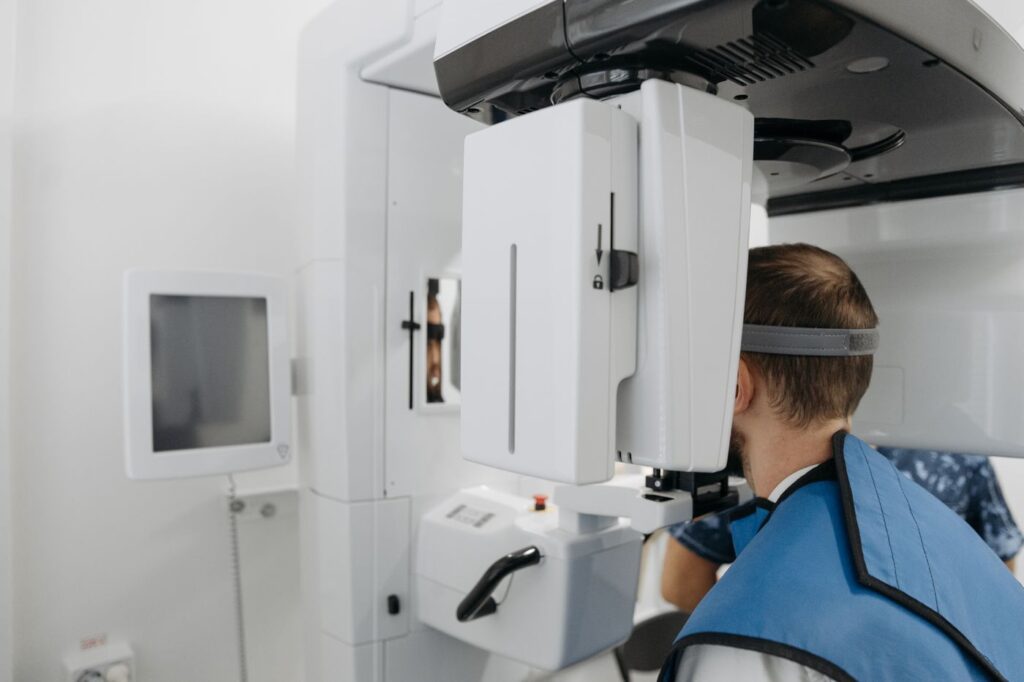
    Cone-beam imaging takes a series of images as the machine rotates, then reconstructs them into a volume that can be viewed in cross-section from any angle.
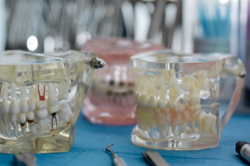
    An extra canal hidden behind another is the classic 3D finding
    An extra canal hidden behind another is the classic 3D finding.
    What each type of imaging shows
    QuestionStandard radiograph3D scan
    How many canals does this tooth have?Often incompleteReliable, including canals hidden behind others
    How far has the infection spread?Underestimates early lesionsShows true extent in three dimensions
    Is there resorption, and how far around?Frequently missedShows position and circumferential spread
    Where is the sinus or nerve relative to the root?ApproximatePrecise — important before surgery
    Is this root fractured?Rarely visibleSometimes visible; still not guaranteed
    Routine diagnosis of a straightforward toothEntirely adequateUnnecessary

    Swipe the table sideways to see every column.

    That last row matters as much as the others. For most teeth a conventional radiograph answers the question completely, and taking a scan adds dose without adding information.

    When it is actually indicated

    The joint position of the endodontic and oral radiology specialties is that scans should be used selectively, guided by the clinical question. In practice that means:
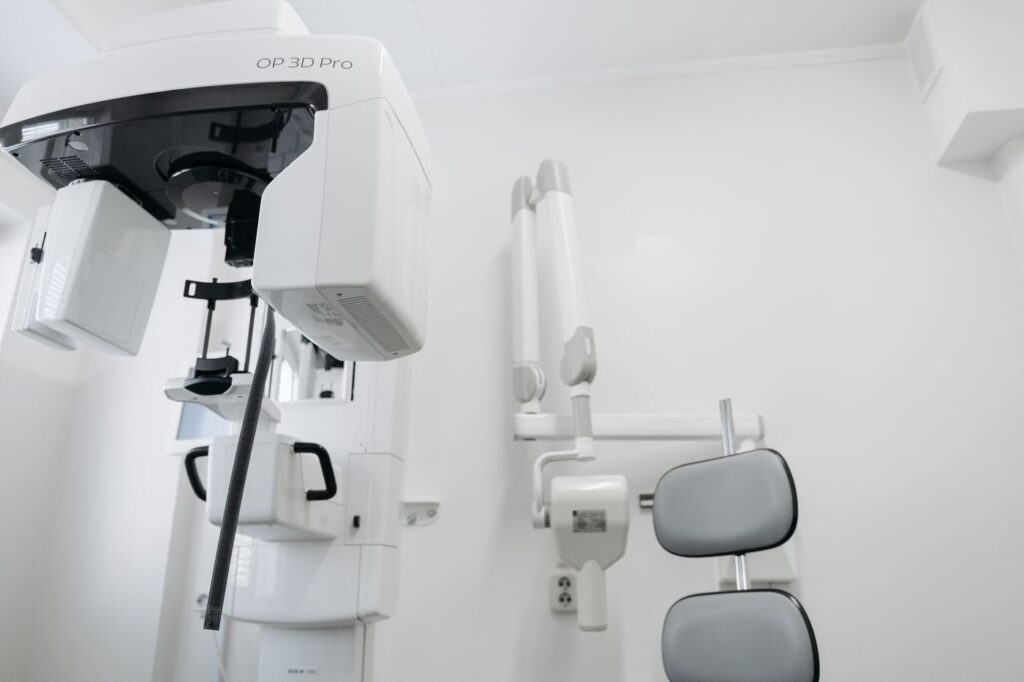
    A limited field scan covers a few teeth at a far smaller dose
    A limited field scan covers a few teeth at a far smaller dose than a full-head study.
    • Retreatment, where the reason the first attempt failed is not visible — see why root canals fail.
    • Suspected extra canals, particularly in upper molars.
    • Suspected resorption, where extent decides whether the tooth is restorable — see how resorption is assessed.
    • Before surgery, to establish exactly where the sinus floor or the nerve to the lip sits relative to the root tip.
    • Dental trauma, where root fractures and displacement are hard to assess on a film.
    • Persistent symptoms with no explanation on conventional imaging.

    What we actually find

    Scans are worth taking only where the finding changes something. In endodontic practice a handful of findings recur.

    Common scan findings and what each changes
    FindingHow often it mattersWhat changes
    An untreated canal in a previously treated toothThe commonest useful findingRetreatment becomes worthwhile instead of extraction
    A lesion larger than the film suggestedFrequentPrognosis and follow-up interval
    Resorption, and how far around the rootOccasional but decisiveWhether the tooth is restorable at all
    Root tip within or against the sinusCommon in upper molarsSurgical approach, and what we warn you about
    Nerve canal close to a lower rootCommon in lower molarsWhether surgery is advisable
    Nothing unexpectedOftenConfirms the original plan — still useful

    Swipe the table sideways to see every column.

    The last row deserves as much weight as the first. A scan that confirms there is nothing hidden is not a wasted scan; it converts a suspicion into a decision, and it is frequently what allows treatment to proceed with confidence rather than caution.

    It also occasionally shows something outside the tooth entirely — sinus disease, or a lesion unrelated to the tooth we were investigating. Where that happens we refer it on rather than treating around it.

    The dose question, answered properly

    A scan is more radiation than a single film. That is worth stating plainly rather than glossing.
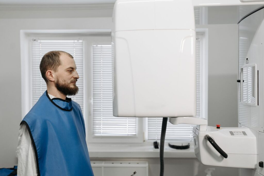
    Scans are taken selectively, never routinely — the ALARA principle
    Scans are taken selectively, never routinely — the ALARA principle.

    The relevant comparison, though, is not scan versus nothing. It is scan versus the several conventional images at different angles that would otherwise be taken trying to answer the same question — and often failing. A limited field-of-view scan is roughly equivalent to two periapical radiographs, and in a complex case it can represent a net reduction.

    The governing principle is ALARA — as low as reasonably achievable. That means the smallest field of view that answers the question, taken only when the answer will change what we do. It also means we do not take one just because the machine is there.

    Where it changes the plan

    Imaging is only worth taking if it can change a decision. In endodontics it regularly does.
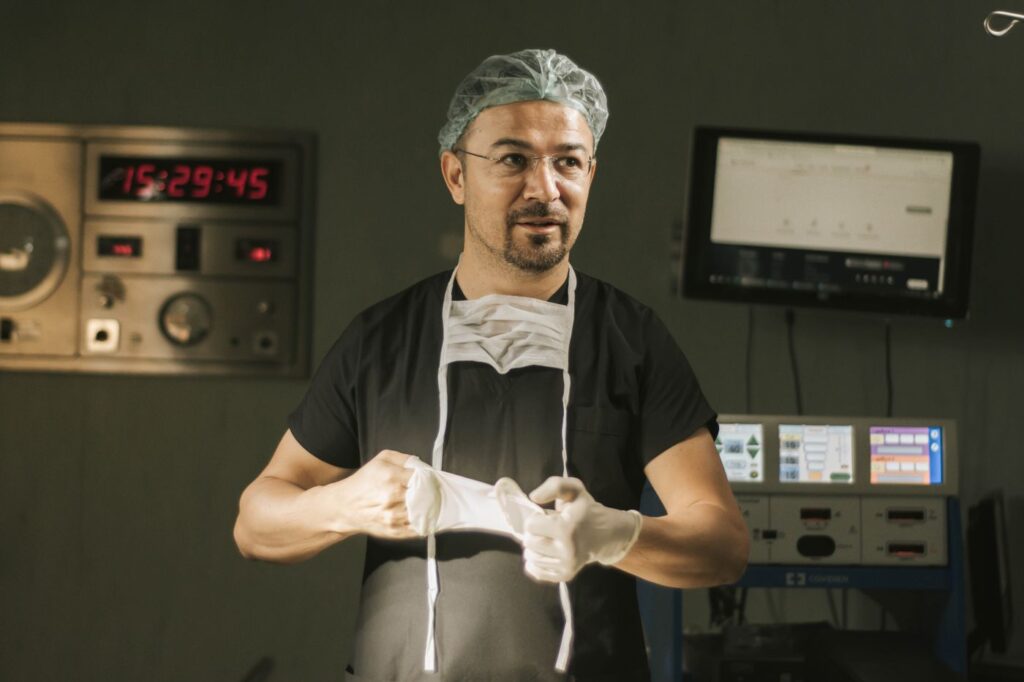
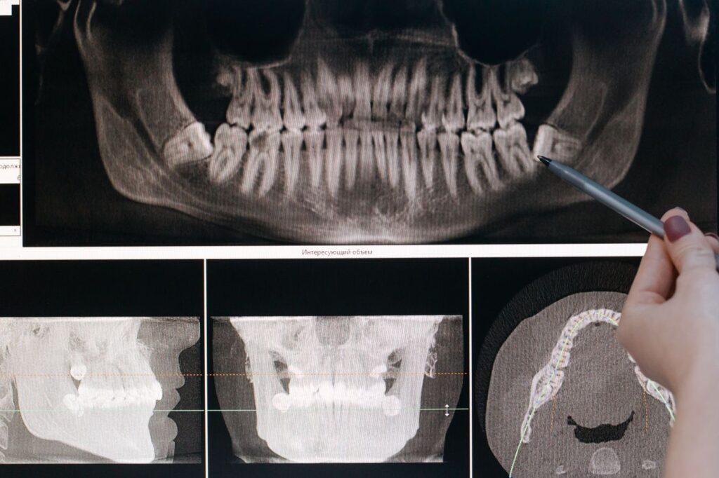
    Before surgery, 3D shows exactly where the sinus and nerve sit
    Before surgery, 3D shows exactly where the sinus and nerve sit.

    A scan showing an untreated canal converts a tooth destined for extraction into one that can be retreated. A scan showing resorption wrapped around most of a root converts an optimistic repair plan into an honest conversation about the alternatives. Before root-end surgery, it turns an estimate about where the nerve runs into a measurement. It also earns its place in heavily calcified teeth, which is why it comes up so often in treatment planning for older patients.

    What it is like to have one

    Undramatic, which is rather the point of mentioning it. You sit or stand with your chin resting on a support while the arm rotates once around your head. Ten to twenty seconds, no enclosed tube, nothing injected, no dye to swallow, and you can be spoken to throughout.
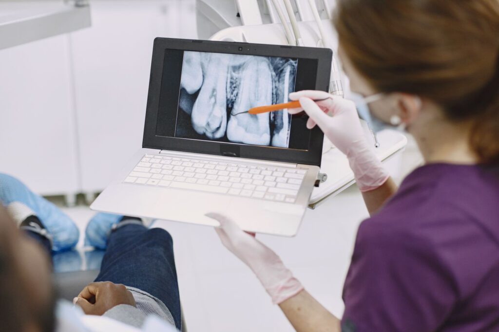
    Better information changes the plan, not just the picture
    Better information changes the plan, not just the picture.

    We review the volume with you where it helps — seeing an extra canal on screen explains a treatment plan faster than any description. Our page on the technology used in the practice covers the rest of the equipment, and the joint position statement on cone-beam imaging in endodontics sets out the professional criteria.

    Frequently asked questions

    Is a 3D scan more radiation than an X-ray?

    Yes, but less than people assume. A limited field-of-view endodontic scan delivers roughly the dose of about two standard periapical radiographs, and it can save taking several conventional images in a complex case.

    Will I need one?

    Probably not. Most root canal treatment is planned from standard radiographs. Scans are taken selectively — for retreatment, suspected extra canals, resorption, trauma, or before surgery.

    What does the scan feel like?

    Nothing. You sit or stand still while the machine rotates around your head for ten to twenty seconds. There is no enclosed tube and no injection.

    Can my dentist see the scan too?

    Yes. Where a scan informs the treatment plan, the findings go into the report we send back to your dentist, along with post-treatment radiographs.

    Have Questions or Ready to Schedule?

    Our team is here to help — call, email, or request an appointment online.
    About this guide. Written for patients of Southeast Endodontics, PC, Charleston, SC. Whether a scan is indicated for your tooth is a clinical judgement made at the consultation, not a routine part of every visit.

  • GentleWave vs. Traditional Root Canal: Cost, Comfort and Results

    GentleWave vs. Traditional Root Canal: Cost, Comfort and Results

    If your endodontist has offered the GentleWave Procedure, the useful question is not whether the technology is impressive — it is whether it changes anything that matters to you as a patient.

    Here is what actually differs between GentleWave and a conventional root canal, what it costs, and the cases where conventional treatment remains the better choice.

    Key takeaways

    • Both approaches do the same job: remove infected pulp tissue, disinfect the canal system, and seal it.
    • The difference is how the canals are cleaned — mechanical files versus a fluid vortex that reaches spaces files cannot.
    • The practical patient benefits are usually fewer appointments and less post-operative soreness.
    • It costs more, mostly because insurers have no separate code for it. It is also not suitable for every tooth.

    The difference in one paragraph

    A conventional root canal cleans the canal by mechanically enlarging it with a sequence of fine files, then rinsing with disinfectant. GentleWave keeps the fluid itself in constant motion using acoustic energy, so the disinfectant reaches the branches and side canals that files physically cannot enter.

    Side-by-side comparison

    GentleWave vs. traditional root canal treatment: what actually differs
    FactorTraditional root canalGentleWave Procedure
    How canals are cleanedSequence of mechanical files, then irrigationFiles used minimally; a circulating fluid vortex does the cleaning
    Reaches side canalsLimited — files only follow the main canalYes — fluid travels into branches files cannot enter
    Tooth structure removedMore, to make room for the filesLess, because the canal need not be widened as much
    Typical appointmentsOften two visitsUsually completed in one
    Post-operative sorenessCommon for a few daysFrequently milder, though not eliminated
    Cost to patientStandard endodontic feeStandard fee plus a technology premium
    Insurance treatmentCovered as normalRoot canal covered; premium usually out of pocket
    Suitable for every toothYesNo — needs enough structure to seal the platform

    Swipe the table sideways to see every column.

    Why cleaning the canal is the hard part

    A molar root canal is not a single tube. It is a branching network with side canals, connecting passages and a fine mesh at the root tip, and the whole system has to be disinfected for treatment to hold.

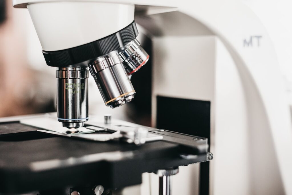
    High magnification lets an endodontist see canal anatomy conventional tools miss
    Magnification reveals canal anatomy that is invisible to the naked eye — and much of it is too fine for any file to enter.

    Files can only follow the main canal. Everything branching off it depends on the irrigant reaching in on its own — which, in still fluid, it does poorly. This is the gap the technology is designed to close, and it is the same reason thorough root canal treatment is a specialist procedure rather than a routine one.

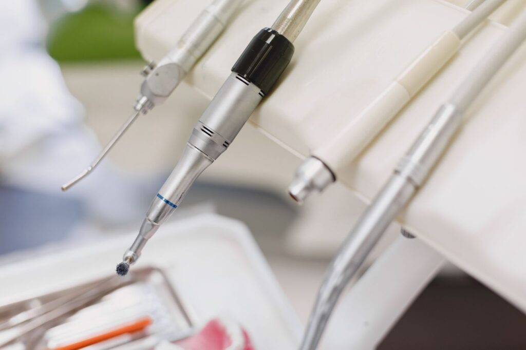
    Conventional root canal treatment relies on mechanical files to shape each canal
    Conventional treatment shapes each canal with a graded sequence of files before disinfecting it.

    How GentleWave works

    A sealed platform is placed over the tooth and filled with disinfecting fluid. The handpiece then generates a vortex with acoustic energy, circulating that fluid throughout the entire canal system at once rather than one canal at a time.

    GentleWave cleans the canal system with a fluid vortex rather than files alone
    Acoustic energy keeps the disinfectant in continuous motion, carrying it into spaces an instrument cannot reach.

    Because the fluid does the cleaning, far less of the canal wall has to be cut away to make room for instruments — which leaves more of your natural tooth intact. That matters years later, since a tooth that keeps more of its own structure is a tooth less likely to fracture under load.

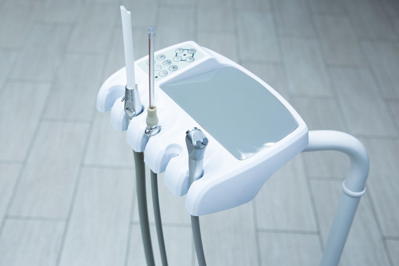
    An operatory equipped for advanced endodontic care
    The system is one part of a wider setup — magnification and 3D imaging do as much for the outcome as the irrigation itself.

    No single piece of equipment carries a case on its own. Our page on the technology used in our Charleston practice covers what else is involved, and the GentleWave treatment overview explains the procedure step by step.

    What it costs, and why

    Expect a premium of a few hundred dollars over a conventional root canal. The reason is administrative rather than clinical: insurers reimburse a root canal under one code and have not created a separate one for this technology, so the difference falls to the patient.

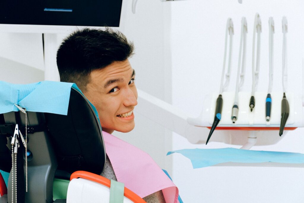
    Fewer appointments means less time away from work and family
    For patients travelling in or taking time off work, collapsing two visits into one is often the deciding factor.
    What you pay, and what insurance typically does with it
    ItemTraditionalGentleWave
    Root canal feeBilled under standard endodontic codeSame code, same fee
    Technology premiumNoneCommonly a few hundred dollars, usually out of pocket
    Crown afterwardsNearly always neededNearly always needed
    Second appointmentOften requiredUsually avoided — saves a day off work

    Swipe the table sideways to see every column.

    Some teeth benefit far more than others. Lower molars often have narrow, curved canals with side branches that files struggle to reach, so a fluid-based clean has real work to do; a straightforward single-canal front tooth gives it much less to improve on. If you are deciding whether the premium is worth paying, the complexity of your particular tooth is a better guide than the technology itself.

    Ask for a written estimate before treatment. Our notes on dental insurance and endodontic coverage and the office policy and fees page set out how we handle this.

    When conventional treatment is the better choice

    The system needs a sealed platform over the tooth to hold the fluid under controlled pressure. Where that seal cannot be achieved reliably, conventional treatment is more predictable:

    Any endodontist recommending it for every single tooth is overselling it. The honest position is that it is an excellent default with a real list of exceptions.

    What it is like as a patient

    You are numb either way, and the appointment runs much as any root canal does. The audible difference is less of the high-pitched drilling associated with files.

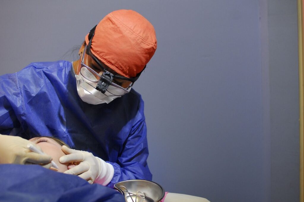
    Most GentleWave cases are completed in a single appointment
    From the chair, the two procedures feel broadly similar — the differences show up afterwards.

    Afterwards, most patients report milder soreness, largely because less instrumentation means less irritation at the root tip. See how to prepare before endodontic treatment and what happens at your first visit. Sonendo publishes its own comparison of standard treatment and the GentleWave Procedure if you want the manufacturer’s detail.

    Frequently asked questions

    How much more does GentleWave cost than a traditional root canal?

    Most practices charge a premium of a few hundred dollars, largely because insurers have not yet created a separate billing code for it. In Sonendo’s own survey, 81% of respondents said they would pay at least $250 more for it.

    Does insurance cover the GentleWave procedure?

    Insurance covers the root canal itself at your usual endodontic rate. The technology premium is typically an out-of-pocket difference rather than a covered item — ask for a written estimate before treatment.

    Is GentleWave painful?

    No more than a conventional root canal, and often less afterwards. The tooth is fully anaesthetised either way. Because less mechanical filing is needed, post-operative soreness is frequently milder.

    Can every tooth be treated with GentleWave?

    Most can, but not all. The system needs a sealed platform to hold the fluid, so teeth with very little structure left, certain fractures, or some retreatment cases are better handled conventionally.

    Have Questions or Ready to Schedule?

    Our team is here to help — call, email, or request an appointment online.
    About this guide. Written for patients of Southeast Endodontics, PC in Charleston, SC. Southeast Endodontics has no financial relationship with Sonendo beyond purchasing the equipment. Costs are general ranges rather than a quote — ask for a written estimate for your specific tooth.