Resorption is one of the few dental conditions where the body dismantles its own tooth structure. It is usually painless, frequently invisible until it is advanced, and almost always found by accident.
Here is what it is, why an old injury decades ago can still be the cause, and what determines whether a resorbing tooth can be kept.
Key takeaways
- Specialised cells break down dentine from either inside the canal or the outer root surface.
- It is typically painless, so it is usually spotted on a radiograph rather than reported as a symptom.
- Past trauma is the commonest trigger, sometimes many years earlier.
- Prognosis depends almost entirely on how early it is found.
What resorption actually is
Your body constantly remodels bone using cells that dissolve hard tissue and cells that rebuild it. Teeth are normally protected from this by their outer layers.
When that protection is damaged — by injury, inflammation or pressure — those dissolving cells can gain access to dentine and start removing it. Nothing rebuilds it, so the process is one-way. In children this is entirely normal and it is how baby teeth loosen; in an adult tooth it is pathological.
Internal and external resorption
| Type | Where it starts | Typical cause | Outlook |
|---|---|---|---|
| Internal | Inside the root canal, working outward | Chronic pulp inflammation after trauma or deep decay | Good if treated before perforation |
| External cervical | At the neck of the root, under the gum | Trauma, orthodontics, older internal bleaching | Depends heavily on how far it has spread |
| External inflammatory | Root surface, driven by infection | Usually follows avulsion or luxation injury | Needs prompt root canal treatment |
| External replacement | Root fuses to bone and is replaced by it | Severe trauma, often a dried-out replanted tooth | Poor — the tooth is gradually lost |
Swipe the table sideways to see every column.
That last row is why how a knocked-out tooth is handled in the first thirty minutes matters so much. A tooth allowed to dry out before replanting frequently develops replacement resorption years later.
Why it is usually found by accident
Resorption has almost no early symptoms. It does not usually cause pain, the tooth stays firm, and in external cervical resorption the lesion is hidden beneath the gum.

The one visible sign, when it appears, is a faint pink area showing through the crown — resorptive tissue is vascular, and its colour shows through thinning enamel. By the time that is visible the lesion is well established.
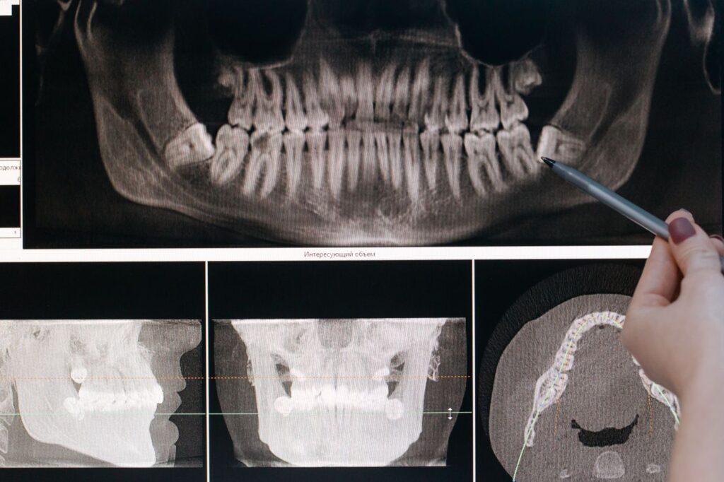
Most cases are therefore found on a radiograph taken for another reason, which is a good argument for keeping routine check-ups even when nothing hurts.
The trauma connection
The single most common history is an injury to that tooth in the past. A childhood fall, a sports impact, a knock that seemed to resolve — and then twenty years later a radiograph shows resorption.

That delay is why the connection is so often missed. Orthodontic movement and older internal bleaching techniques are also associated with it, and chronic inflammation around the tooth can contribute. In a meaningful proportion of cases no cause is ever established.
The signs, such as they are
Resorption is defined more by the absence of symptoms than the presence of them, which is why the list below is short and mostly late.
| What is noticed | What it suggests | Stage |
|---|---|---|
| Nothing at all — found on a routine film | Any type | Early, and the best case |
| A pink patch showing through the crown | Internal, or advanced cervical | Established |
| A notch or roughness felt at the gumline | External cervical | Established |
| Gum bleeding or overgrowth at one spot | External cervical with tissue ingrowth | Established |
| Sensitivity or ache in a previously injured tooth | Inflammatory, pulp becoming involved | Later |
| Tooth feels solid but sounds dull on tapping | Replacement resorption | Advanced |
Swipe the table sideways to see every column.
That last one is worth explaining. A tooth fusing to bone loses the slight cushioning of the ligament, so tapping it produces a higher, harder note than its neighbours. It is a small sign with a serious meaning.
Why 3D imaging changed the diagnosis
A standard radiograph flattens a three-dimensional root into one image. A resorptive defect on the cheek or tongue side of a root is often hidden behind the root itself.
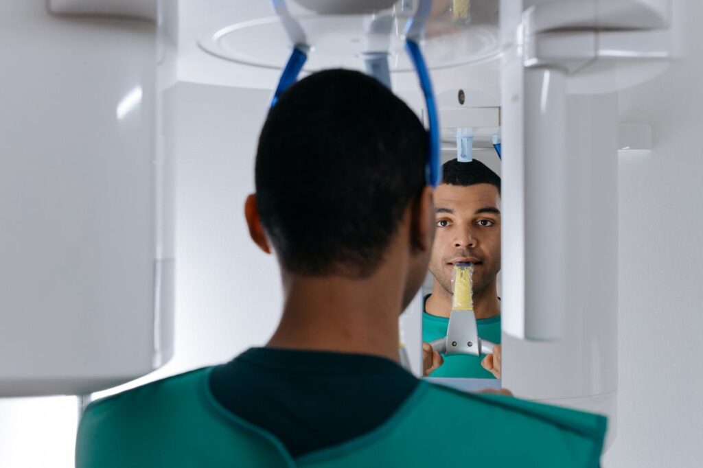
Cone-beam imaging shows the root in cross-section, which reveals both whether resorption is present and how far around the root it has travelled. That second question decides treatment, so a case that looked borderline on a film often becomes clear-cut on a scan — see what a 3D scan shows that an X-ray cannot.
What treatment involves
The principle is straightforward: remove the tissue driving the process and seal the defect so it cannot continue.
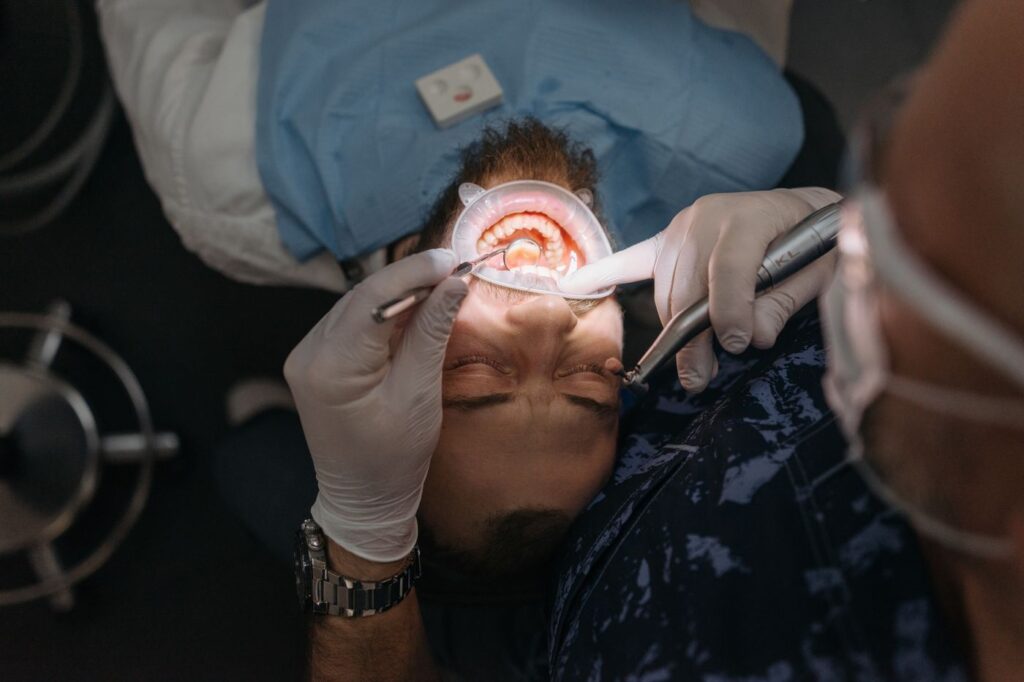
For internal resorption that means root canal treatment, since the process is driven from within the canal. For external cervical resorption, the lesion is usually approached from outside, cleaned out and repaired with a biocompatible material — sometimes with root canal treatment as well, sometimes without if the pulp is uninvolved.
Extensive lesions that have spread circumferentially around the root, or reached deep below the bone level, are frequently not restorable. That is an honest limitation rather than a failure of technique.
Monitoring, and why it is worth it
Not every resorptive lesion needs immediate intervention. Small, stable, symptomless lesions are sometimes monitored with periodic radiographs rather than treated, particularly where treatment would itself be destructive.

What that requires is actually attending the reviews. Resorption is slow but progressive, and the difference between a repairable defect and an unrestorable one is often a couple of years of not being looked at.
If you have had a significant injury to a front tooth at any point, mention it — it changes what we look for. The American Association of Endodontists’ guide to differentiating resorption covers the classification clinically.
Frequently asked questions
What causes tooth resorption?
Most often a past injury — sometimes decades earlier. Orthodontic movement, internal bleaching using older techniques, and chronic gum inflammation are also associated with it. In a proportion of cases no cause is ever identified.
Does it hurt?
Usually not, which is the problem. External cervical resorption in particular is painless and hidden under the gum, so it is often found on a routine radiograph long after it started.
Can a resorbing tooth be saved?
Often, if it is caught early. Small, accessible lesions can be cleaned out and repaired. Extensive lesions that have spread around or deep into the root have a poor outlook and may need extraction.
Is it contagious to other teeth?
No. It is a localised process affecting one tooth, not an infection that spreads. Having it in one tooth does not mean others will develop it, though a shared cause such as past trauma can affect neighbouring teeth.
Have Questions or Ready to Schedule?
Our team is here to help — call, email, or request an appointment online.